Gastric Case 2

Prof. Yip Hon Chi
Department of Surgery, Prince of Wales Hospital,
The Chinese University of Hong Kong, Hong Kong
Scope: GIF-XZ1200
Case: Recurrent gastric dysplasia
Organ: Gastric antrum
Patient information: F, 70s
Medical history: Gastric antral intramucosal adenocarcinoma with curative ESD 16 years ago. Underlying Immune Thrombocytopenia Purpura on steroid (Platelet count ~70 x 109L)
1. WLI observation
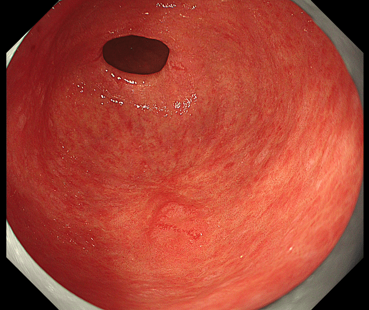
ESD scar is vaguely seen at the greater curve side of antrum, with a slight elevated area proximal to the scar.
2. TXI observation
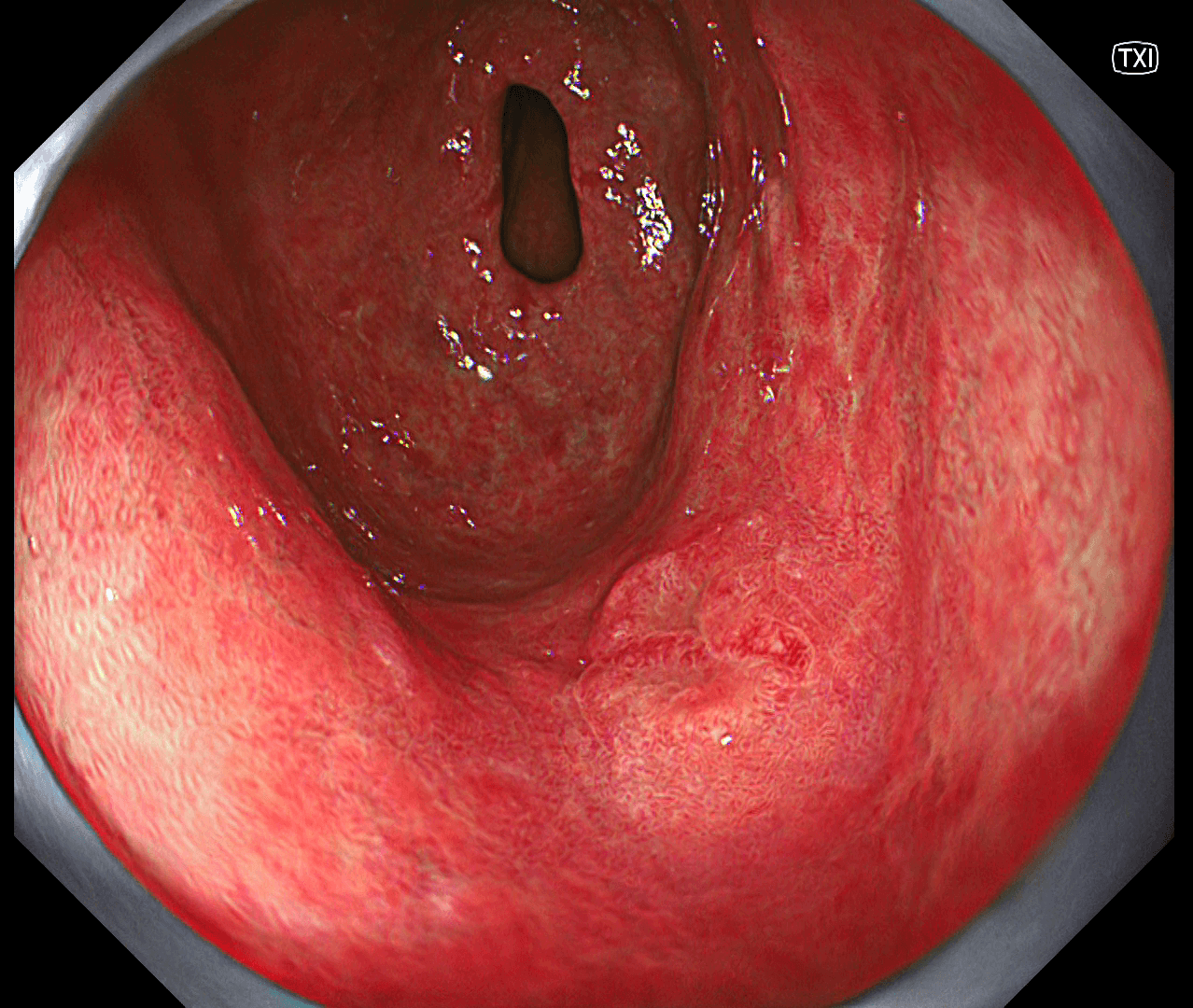
Upon TXI assessment, the boundary of the lesion is clearly seen with irregular shape as well as color, suspicious of neoplastic lesion.
3. NBI-magnification observation
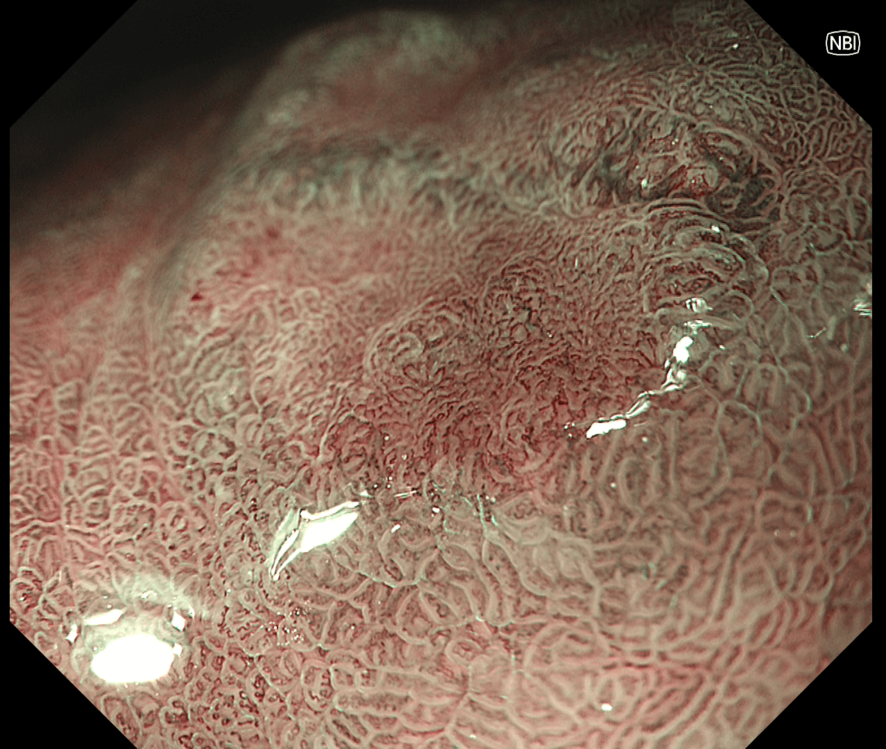
NBI with magnification demonstrated clear demarcation line (DL), irregular microsurface(MS) and microvascular pattern(MV), diagnostic of early gastric cancer.
Case video
Overall Comment
This case demonstrated the utility of the new image processing system in endoscopic diagnosis of early gastric cancer. The patient had a previous early gastric cancer with ESD before. During diagnostic WLI, the ESD scar could be visualized vaguely at the antrum, and a slightly erythematous area was seen proximal to the lesion. Using the TXI mode, the corresponding lesion was greatly enhanced with identification of a clearly demarcated and elevated lesion with irregular colour and morphology. Under NBI with magnification, the lesion margin was assessed clearly with demarcation line and irregular microsurface and microvascular pattern. Based on endoscopic criteria, early recurrent cancer of stomach is highly suspicious, which was subsequently confirmed on biopsy and repeated curative ESD was performed.
* Specifications, design and accessories are subject to change without any notice or obligation on the part of the manufacturer.
Prof. Stefan Seewald Case 3: Multiple gastric polyps (Benign)
Dr. D Nageshwar Reddy
Dr. Hardik Rughwani
- Keyword
- Content Type


