
Takashi Kawai, MD, PhD
Department of Gastroenterological Endoscopy
Tokyo Medical University Hospital, Japan
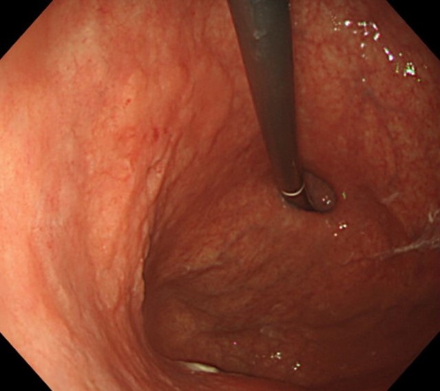
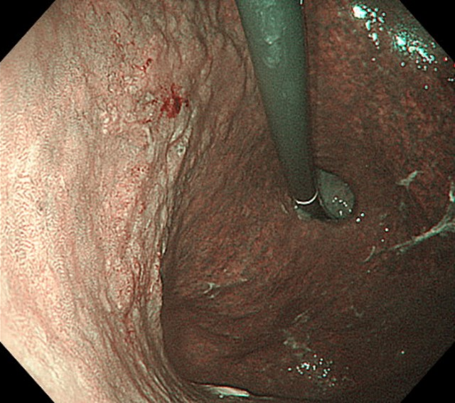
Scope : GIF-1200N / EVIS X1
Case : Early gastric cancer (tub2>tub1, 0-IIa+IIc, pT1b2(SM2), UL(-), ly0, v0)
Organ : Posterior wall of the greater curvature of the upper body of the stomach
Patient Information : M, 70s
Medical History : Nothing relevant
WLI
NBI
Overall comment
Under white light, it was observed that the lesion extended as a low, flat elevation that was mostly the same color as the surrounding tissue with the addition of some paler colors. A clear boundary with the surrounding tissue could be seen and the surface appeared irregular and uneven. Consequently, it was diagnosed as gastric cancer. Narrow Band Imaging (NBI) clarified the demarcation lines and highlighted the irregular uneven micro-surface structure and abnormal dot-like vascular changes on the flat elevated surface. NBI observation was conducted in mid to far distance and the lesion could be clearly observed all the way to the margin thanks to the Brightness Adjustment Imaging with Maintenance of Contrast (BAI-MAC) function.
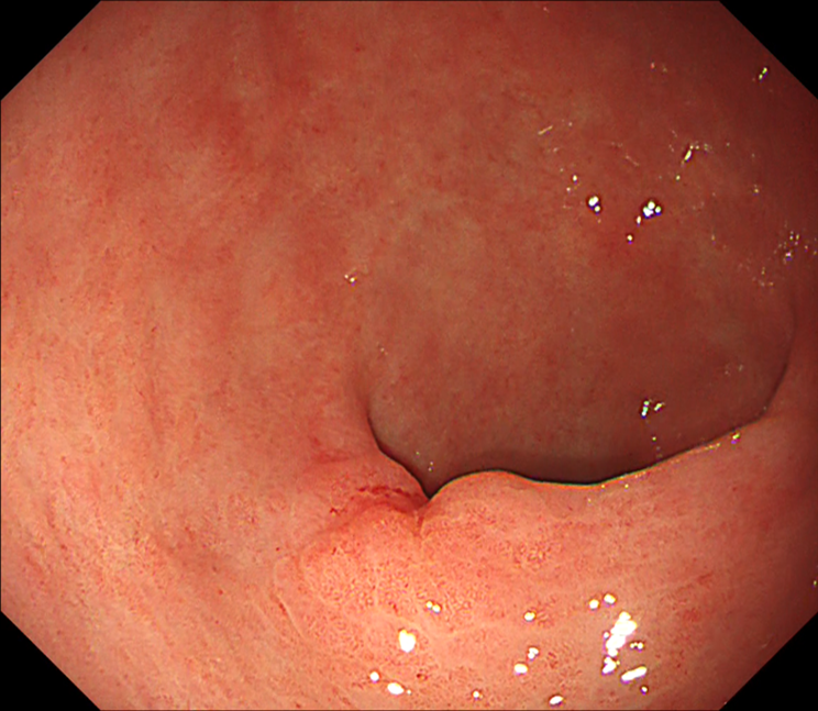
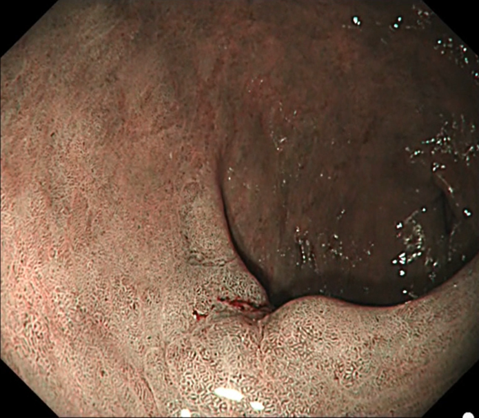
Scope : GIF-1200N / CV290
Case : Early gastric cancer (tub1>tub2, 0-IIc, pT1a(M), UL(-), ly0, v0)
Organ : Greater curvature of the antrum
Patient Information : M, 70s
Medical History : Nothing relevant
WLI
NBI
Overall comment
Under white light, the lesion appeared to be depressed and the boundary with the surrounding tissue was clearly marked. The mucosa in the depression was reddish, which made it possible to diagnose the lesion as gastric cancer. Narrow Band Imaging (NBI) revealed demarcation lines and highlighted the irregular uneven micro-surface structure and abnormal dot-like vascular changes on the flat elevation.
* Specifications, design and accessories are subject to change without any notice or obligation on the part of the manufacturer
Dr. Kunihisa Uchita Case 12: Early gastric cancer (por2>sig>tub2, 0-IIc, pT1a(M), UL(-), ly0, v0)
Takashi Kawai, MD, PhD
- Keyword
- Content Type


