Esophageal Cases 8

Dr. Khanh Do-Cong Pham, FASGE, FESGE
Department of Medicine
Haukeland University Hospital
Bergen, Norway
Scope: GIF-EZ1500, GIF-H190N
Case: Eosinophilic esophagitis (EoE)
Organ: Esophagus
Patient Information: M, 30s
Medical History: More than 10 years history of intermittent dysphagia
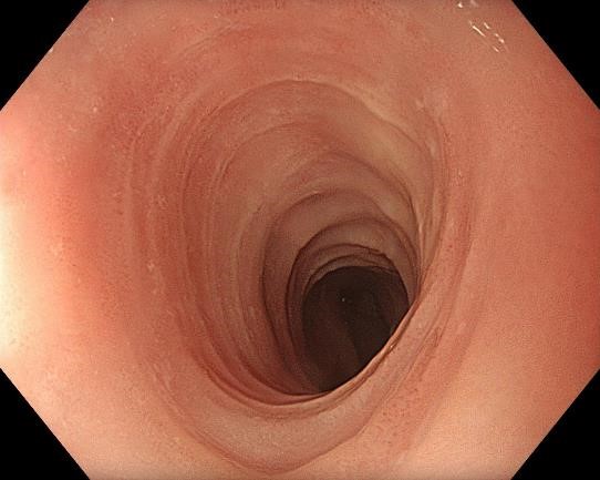
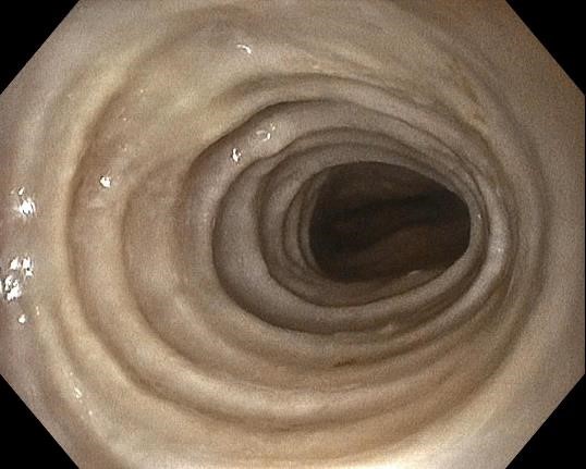
1. EoE in WLI

The mucosa appears normal in the proximal esophagus in white light
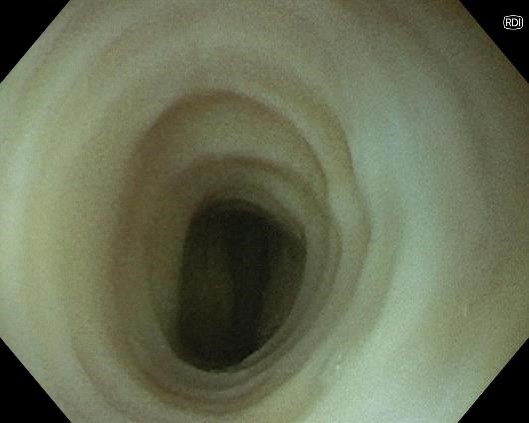
2. EoE with TXI mode 1
The mucosa appears normal in the proximal esophagus with TXI
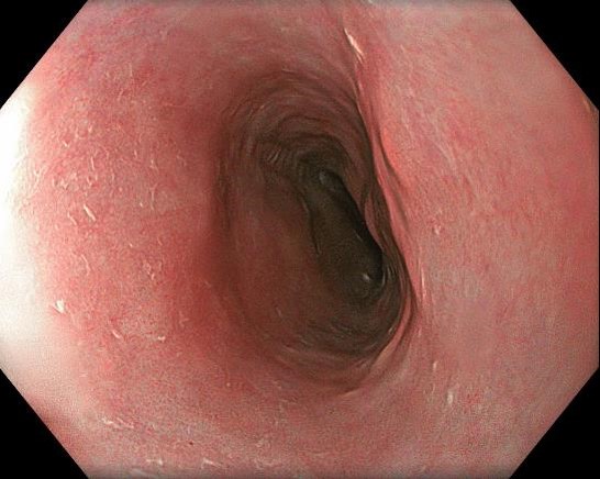
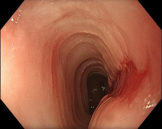
3. EoE stricture a
Stricture in the mid-esophagus.
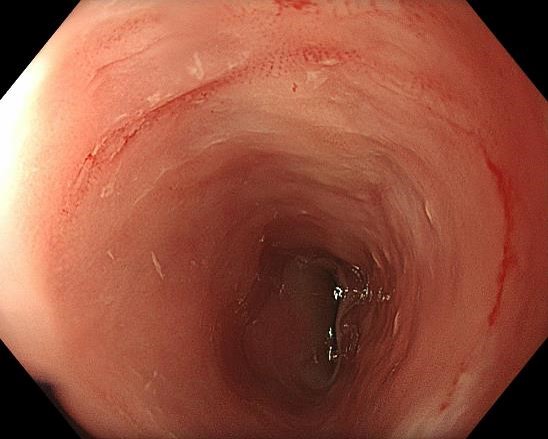
4. EoE Stricture b

Stricture in the mid-esophagus not passable with a standard gastroscope
5. EoE stricture with the ultra-slim endoscope in WLI
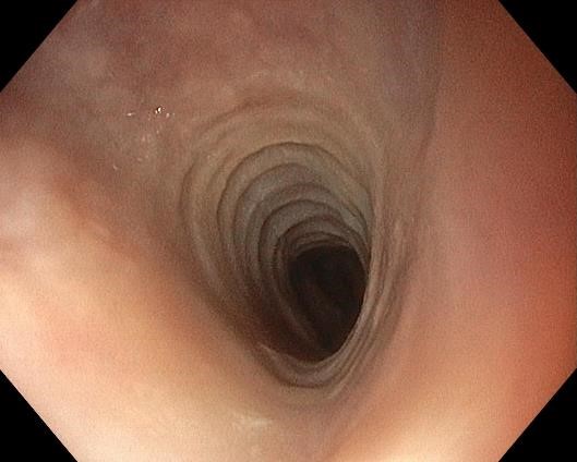
Proximal to the stricture with the ultra-slim endoscope (GIF-H190N) in white light
6. EoE stricture with the ultra-slim endoscope in NBI
Inside the stricture with the ultra slim endoscope (GIF-H190N) with NBI
7. EoE stricture with the ultra-slim endoscope in TXI mode 1
Inside the stricture with an ultra slim endoscope (GIF-H190N) with TXI
8. EoE stricture with the ultra-slim endoscope in RDI mode
Inside the stricture with an ultra slim endoscope (GIF-H190N) with RDI
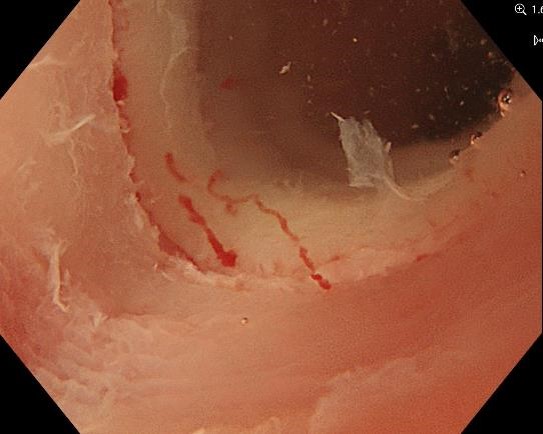
9. Mucosal tear after dilatation
The stricture after dilatation to 12mm. Mucosal trear can be seen, but no perforation.
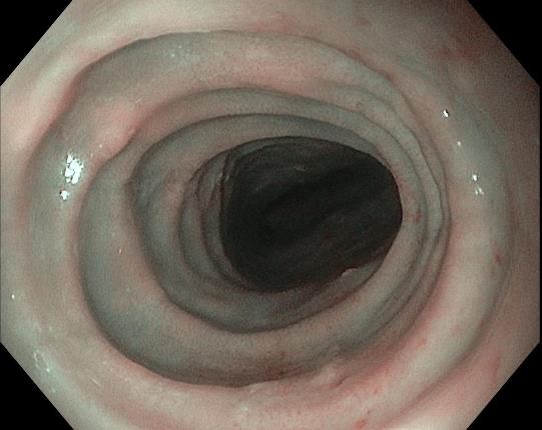
10. The stricture after dilatation
Passing the stricture with the GIF-EZ1500 gastroscope
11. The stricture after dilatation in TXI mode 1 and Near Focus

Details of the stricture can be seen underwater, Near Focus, and TXI
12. The stricture after dilatation in Near Focus and Electric zoom
Details of the stricture seen underwater, Near Focus, and x1.6 electric zoom.
Case video
The GIF-EZ1500 gastroscope was used initially to examine the esophagus. A stricture was found in the mid-esophagus, preventing the further insertion. The mucosa proximally to the stricture was normal. We changed to an ultra-slim endoscope for inspection, passed the stricture, and dilated it to 12 mm. Repeated gastroscopy with the GIF-EZ1500 gastroscope could pass the stricture. The video demonstrates the different optical modes on the EVIS X1 with the different endoscopes.
* Specifications, design and accessories are subject to change without any notice or obligation on the part of the manufacturer
Haruhiro Inoue, MD, PhD Case 9: Barrett esophagus inspection
Dr. Roos Pouw
- Keyword
- Content Type


