Esophageal Case 4

Prof. Stefan Seewald
GastroZentrum Hirslanden, Zurich
Scope:GIF-EZ1500
Case: Esophageal papilloma
Organ: Esophagus
Patient information: F, 67
Medical history: Heartburn
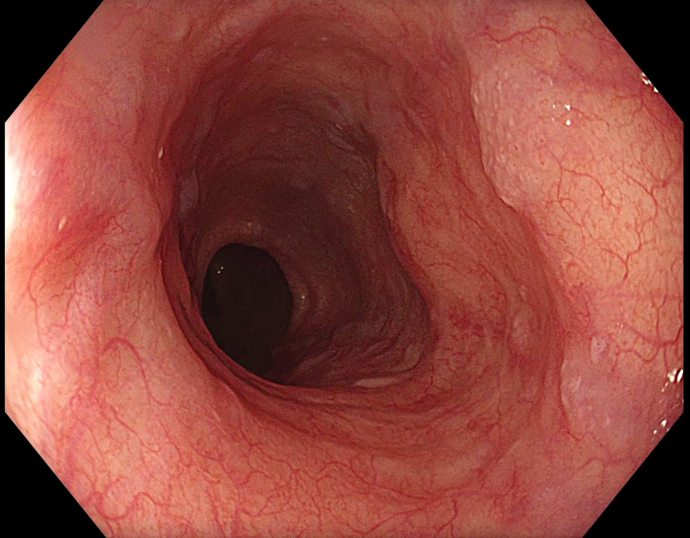
1. WLI
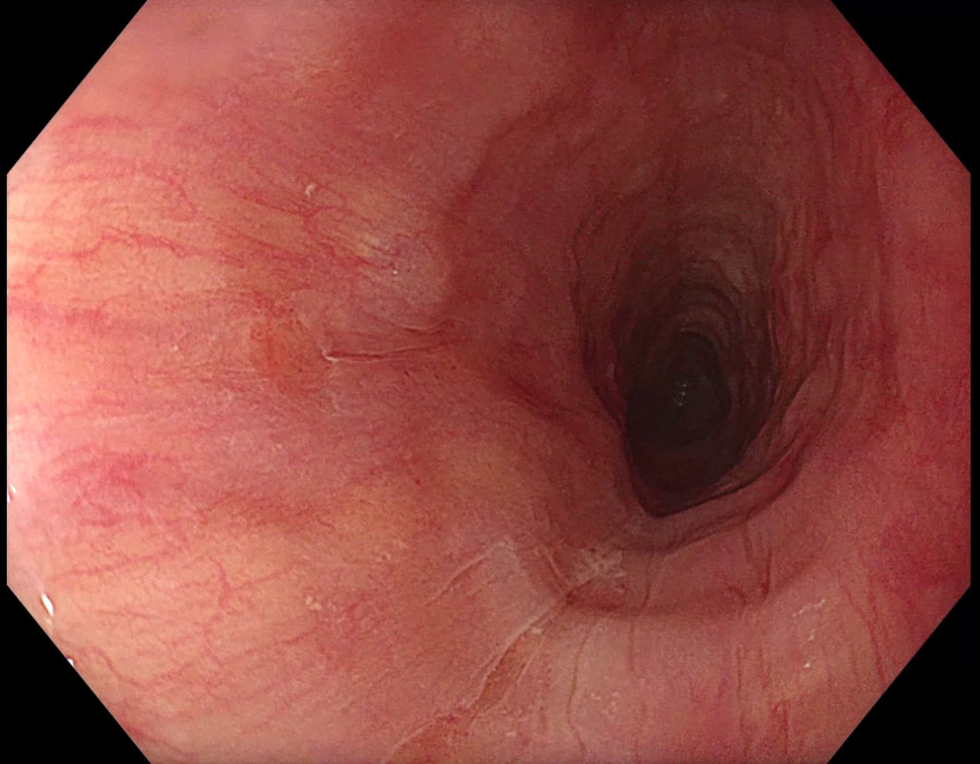
A very small reddish area at 9 o’clock appears suspicious.
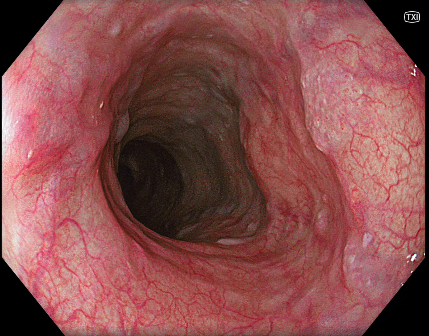
2. TXI
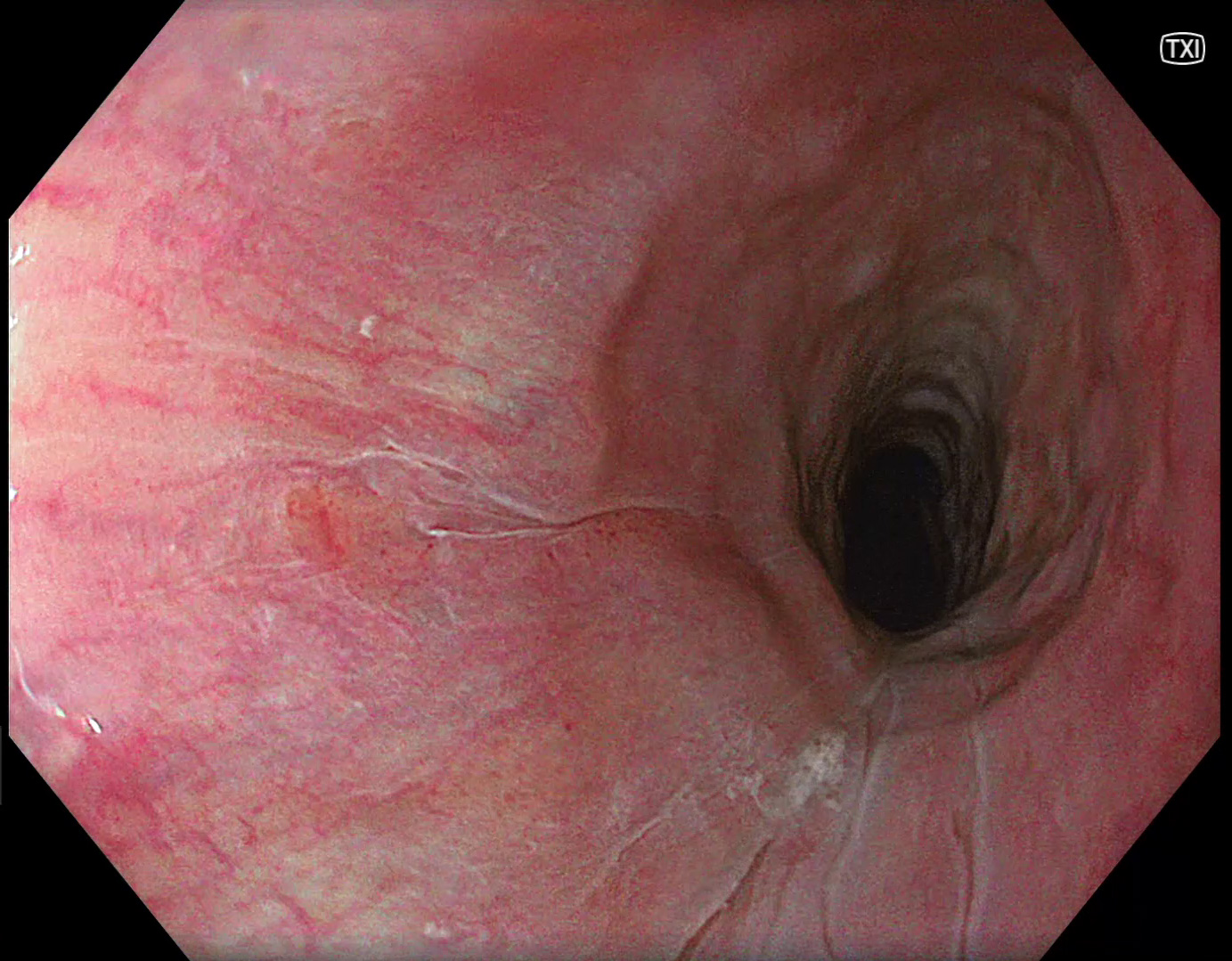
Using TXI, the reddish area is becoming more prominent.
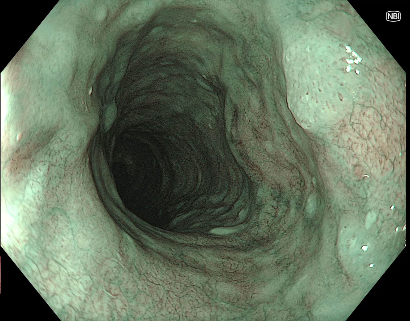
3. NBI
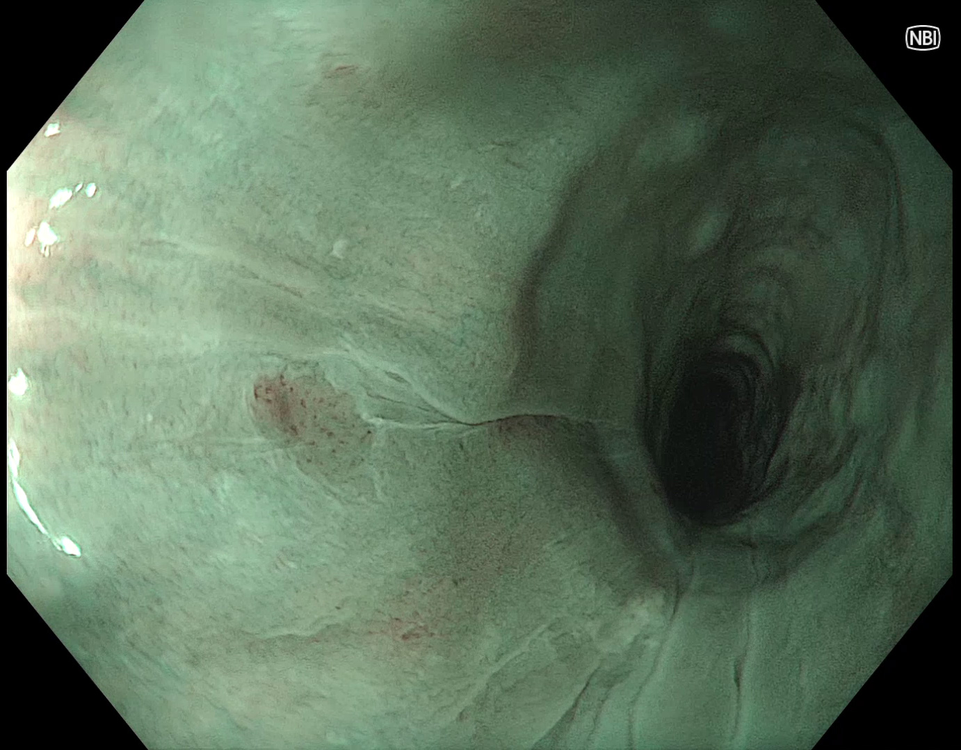
NBI reveals a small brownish area that requires further magnification.
4. Near focus with NBI
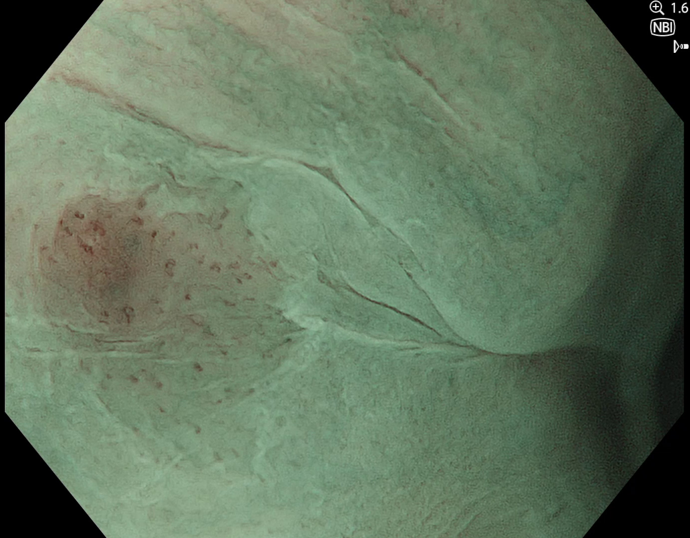
Enlarged IPCLs with brownish background mucosa can be seen.
5. WLI
In the middle esophagus, further papillomas can be easily identified by WLI.
6. TXI
TXI is enhancing the distinct texture component of these lesions.
7. NBI
NBI demonstrates no pathological IPCLs.
8. WLI
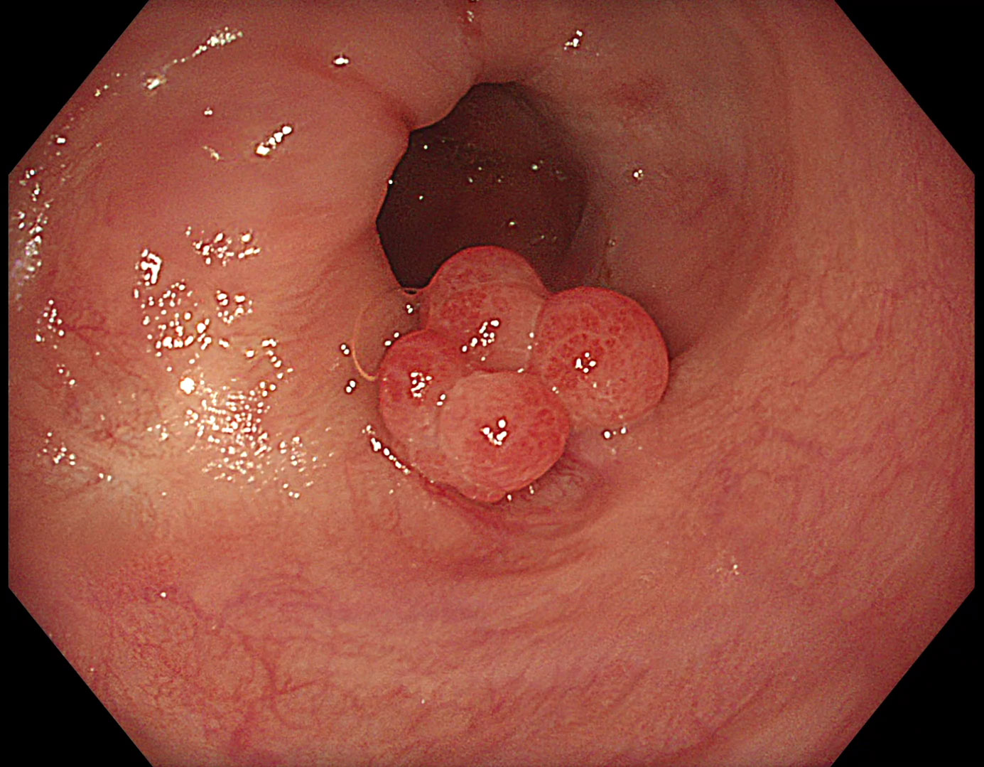
In the lower esophagus, multiple papillomas can be identified.
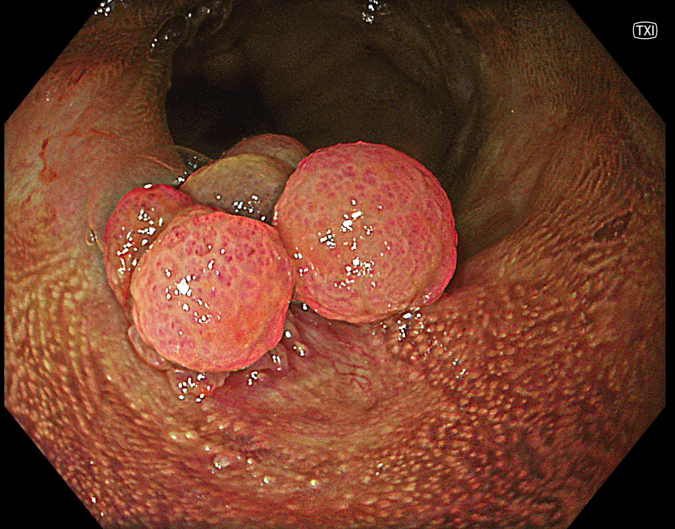
9. TXI
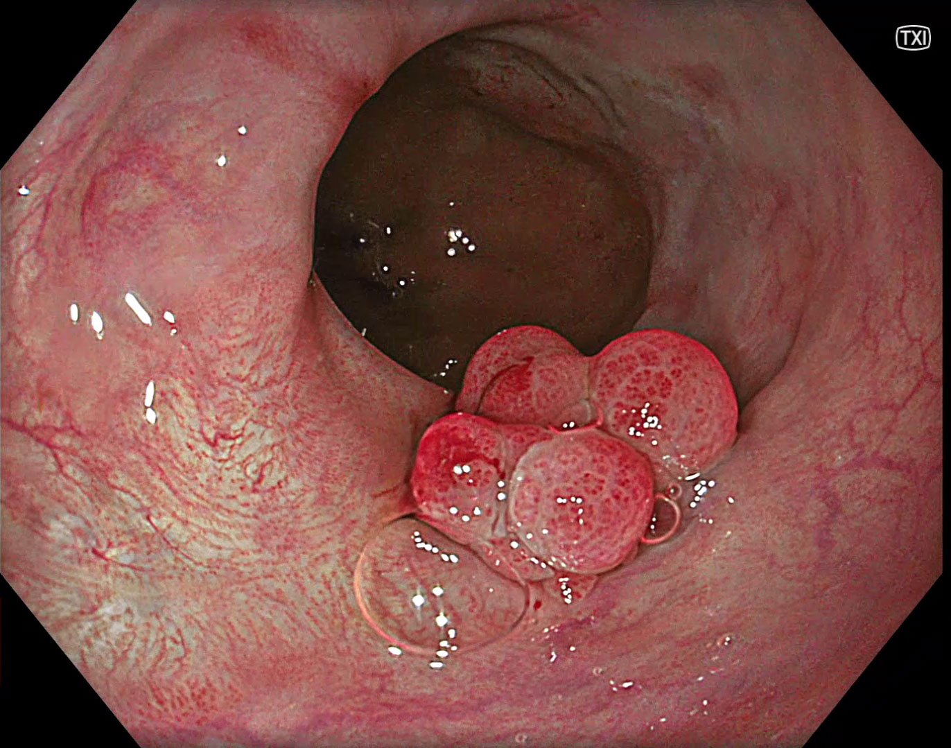
Under TXI, the inflammatory aspect is enhanced by increased color contrast.
10. NBI
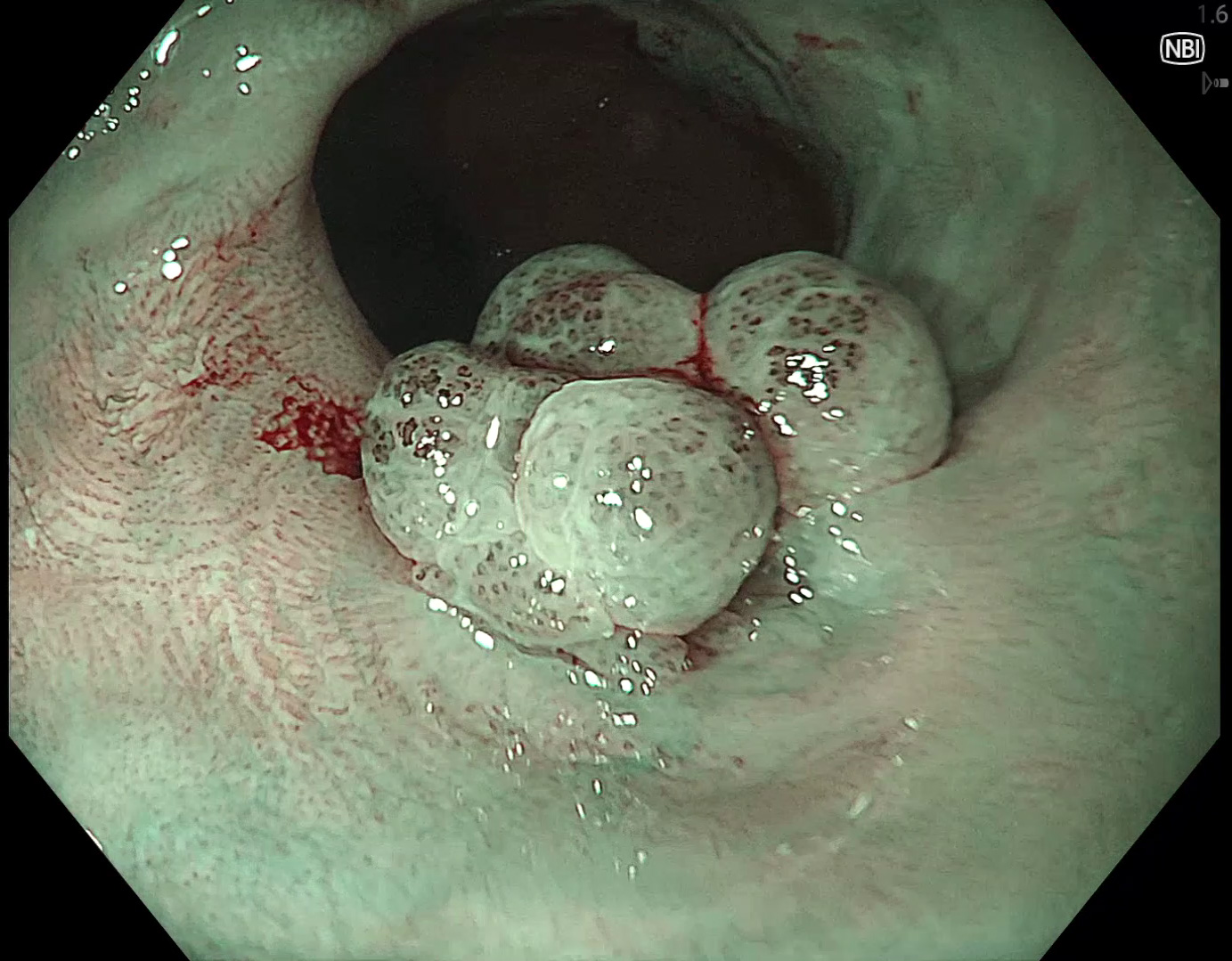
Inflammatory changes around the papilloma can be seen by dilatated IPCLs.
11. Near focus with NBI
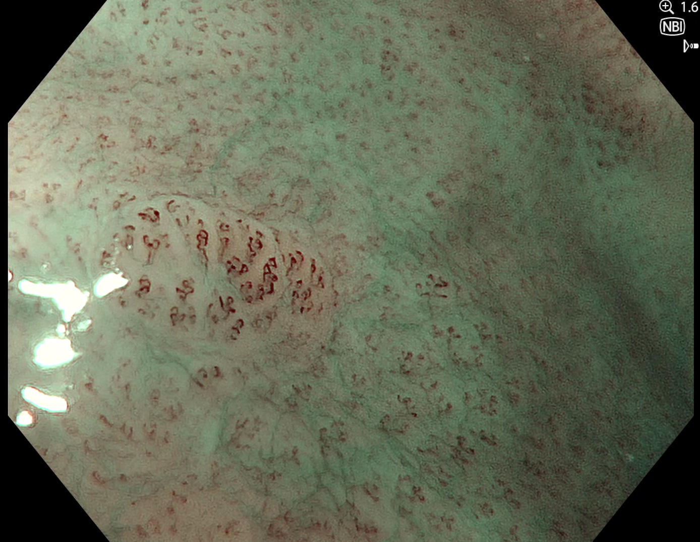
There are additional subtle lesions with IPCL dilatations.
12. Lugol staining and TXI
Lugol staining and TXI show no typical Lugol voiding behaviour.
Case video
* Specifications, design and accessories are subject to change without any notice or obligation on the part of the manufacturer.
Prof. Stefan Seewald Case 5: Early adenocarcinoma in Barrett’s esophagus
Prof. Rajvinder Singh
- Keyword


