Gastric Case 1

Prof. Stefan Seewald
GastroZentrum Hirslanden, Zurich
Scope:GIF-EZ1500
Case: Intramucosal Gastric Carcinoma (AEG Siewert Type III)
Organ: Stomach
Patient information:M, 70
Medical history: Incidental finding
1. WLI
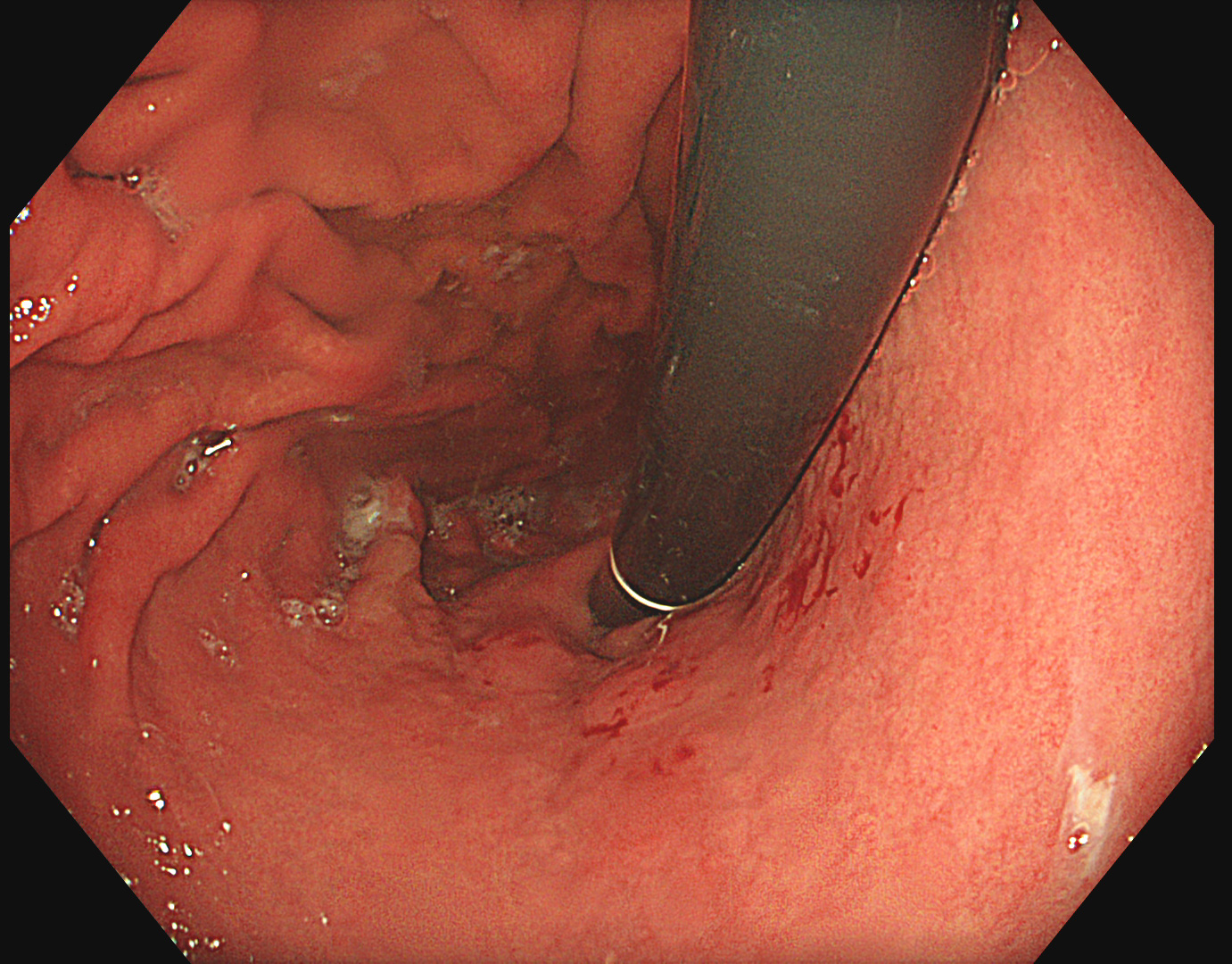
Retroflex assessment of the fundus in white light reveals a suspicious area of patchy appearance spreading from 1-5 o’clock.
2. TXI
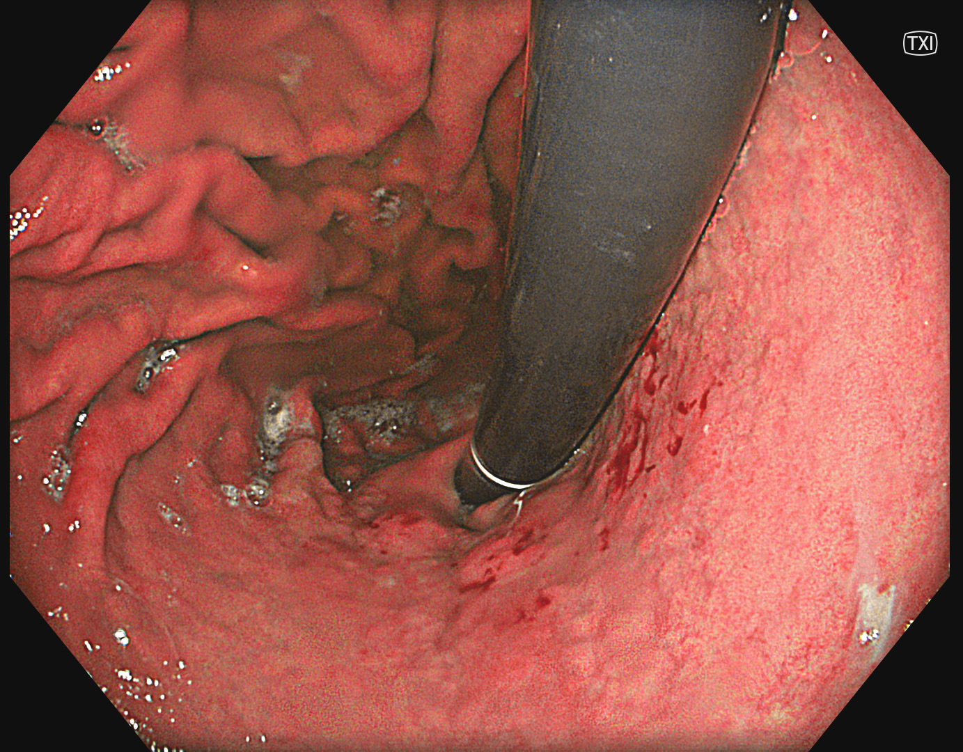
TXI is enhancing the color contrast and structure between the suspicious area and the surrounding mucosa.
3. NBI
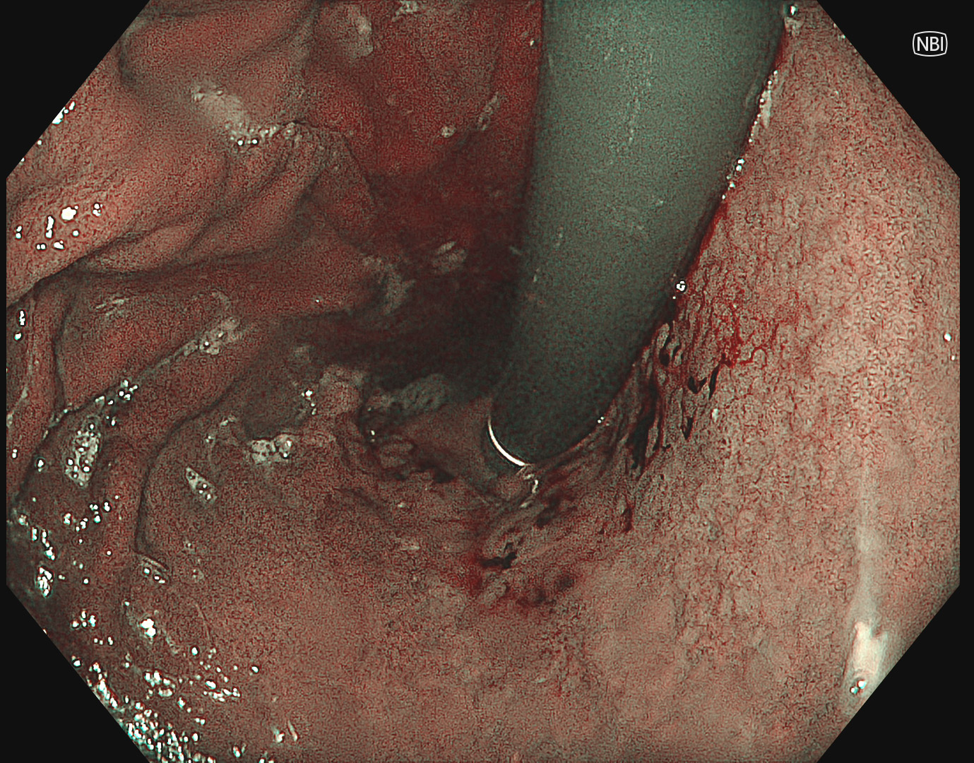
NBI is bright enough but for characterization near focus with NBI is necessary.
4. Near Focus with NBI
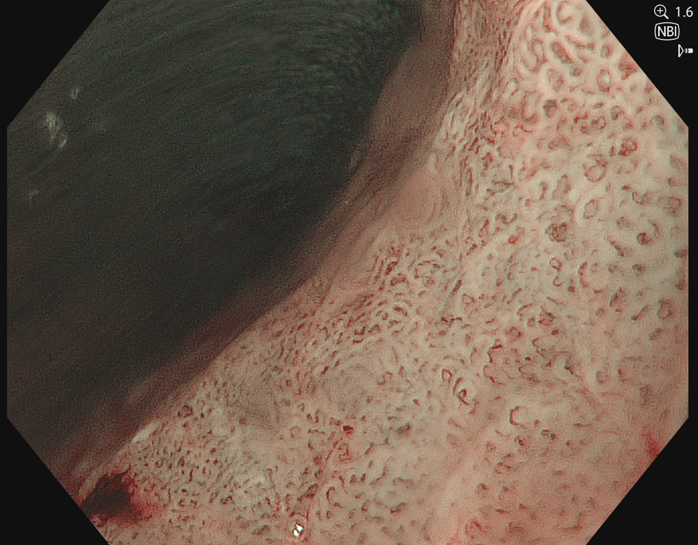
Near Focus with NBI reveals irregular VS pattern suggesting early gastric carcinoma.
5. TXI with indigocarmine
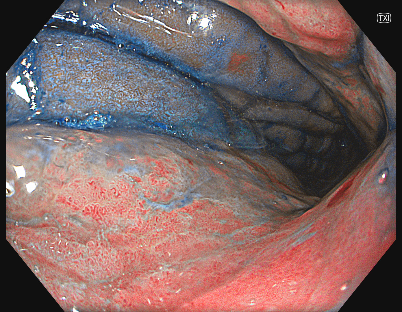
The contrast effect of indigocarmine is enhanced by TXI and improves the delineation.
6. TXI with indigocarmine
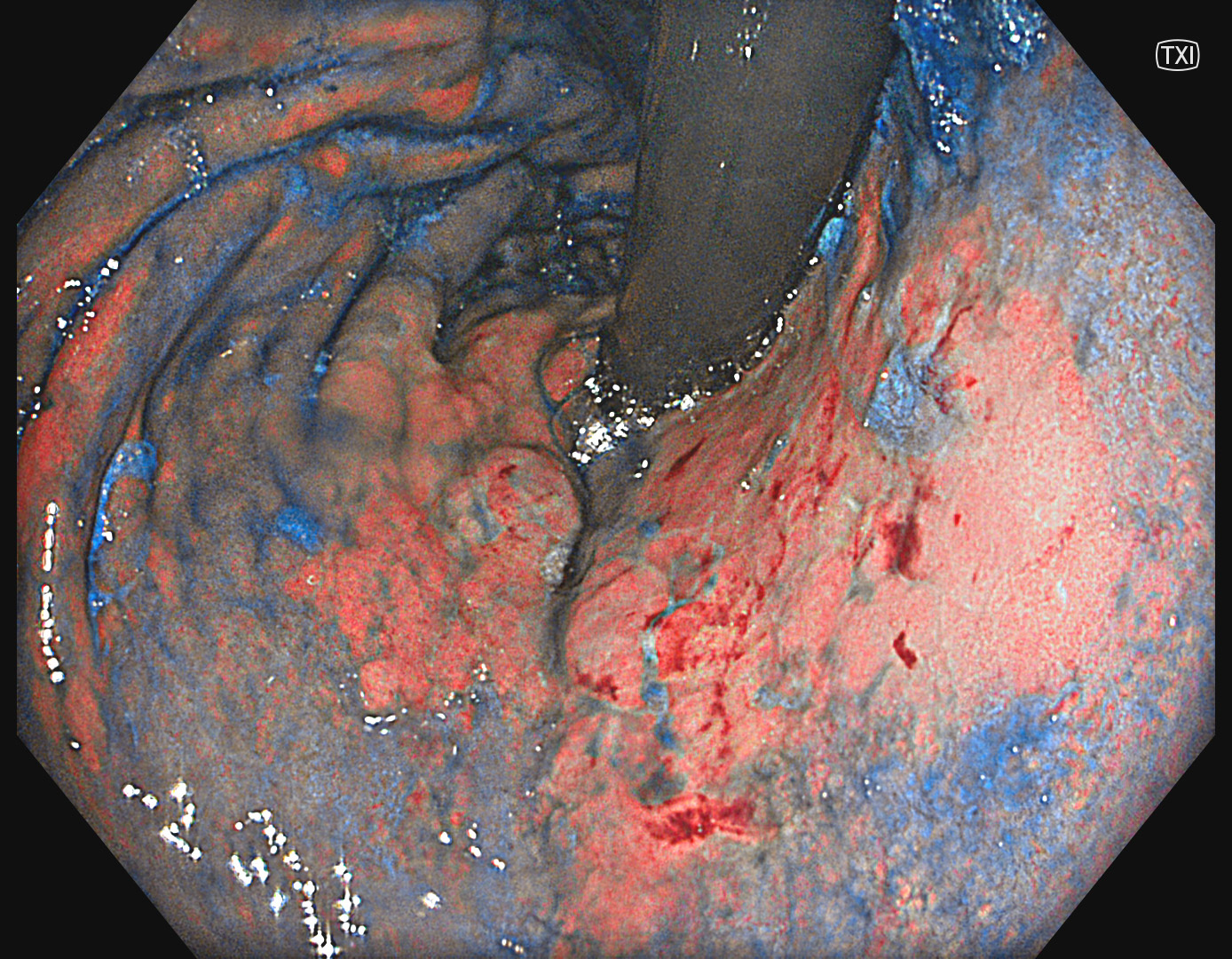
The extent of the lesion can be nicely identified by TXI in combination with indigocarmine.
Overall Comment
The case illustrates the value of TXI in detection and delineation of early gastric carcinomas. Especially the delineation was difficult due to indistinct borders. The combination of TXI and indigocarmine enabled the strong color contrast and delineation of the lesion. NBI remains the first-hand modality for optical diagnosis.
* Specifications, design and accessories are subject to change without any notice or obligation on the part of the manufacturer.
- Content Type


