Case4 – Persistent NSCLC
Author:
Felix Herth, MD and Ralf Eberhardt, MD, Thoraxklinik, University of Heidelberg, Germany
Source:
DVD-ROM ‘Endoscopic Ultrasound – Diagnostics and Staging of Lung Cancer’, Olympus Europa SE & Co. KG, 2013
Patient History
Male, 68 years. Patient with a history of NSCLC (squamous cell lung cancer) stage II B (T2N1M0) in 2008.
Treatment: surgery and adjuvant chemotherapy.
3 months interval routine follow-up with CT. CT shows enlarged lymph node in station 4R in 2010.
Suspicion of right sided mediastinal tumour.
Bronchoscopy
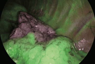
RUL stump shows an area with a different colour compared to the surrounding tissue.
Investigation of the area under Narrow Band Imaging (NBI) reveals concentration of vessels with suspicious tortured structure. Forceps biopsy.
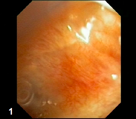
5 mm lesion in the upper area of LN station 4R.
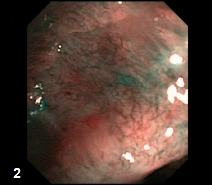
Endobronchial Ultrasound
Echopoor lesion of 14×12 mm close to the superior vena cava (LN 4R).
4
Pathology
Specimen from lymph node station 4R positive for squamous cell carcinoma (NSCLC).
Diagnosis
Local lymph node metastasis of persistent NSCLC.
Therapy
Radiation therapy.
- Content Type