Dr. Vinay Dhir
IDL Care S.L. Raheja Hospital, India
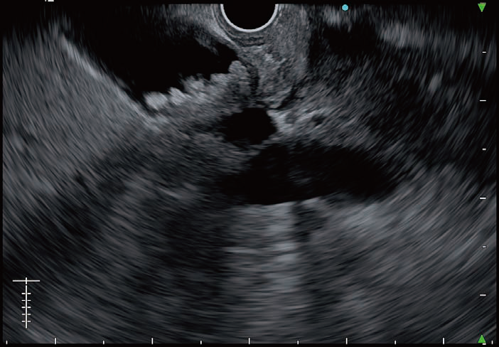
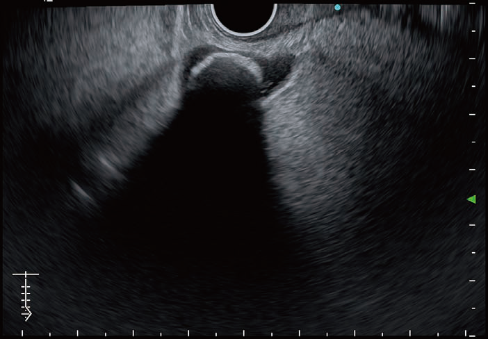
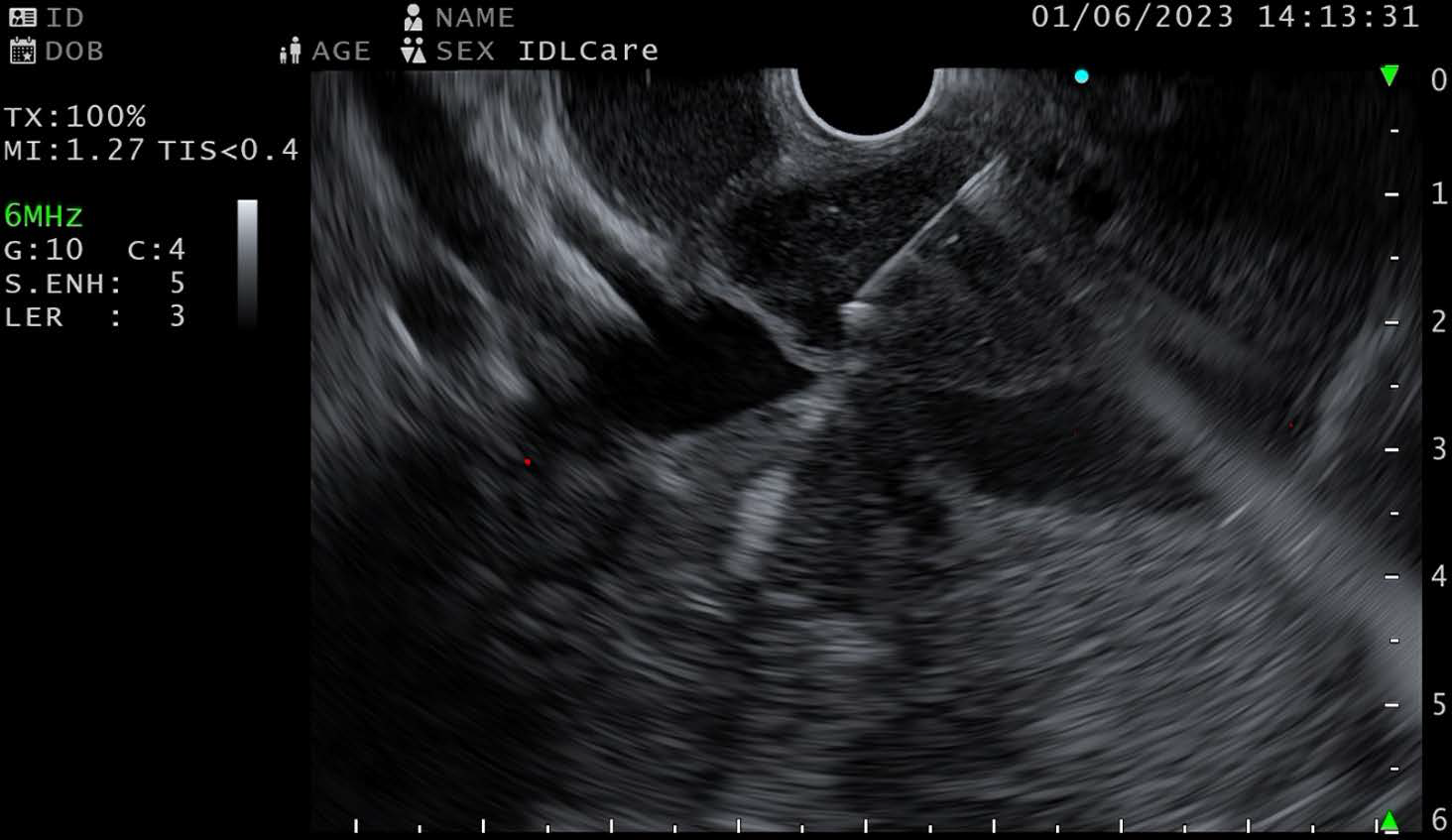
What is your impression of B-mode image of EU-ME3 ?
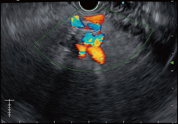
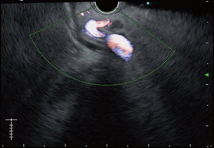
The overall image quality is superior to EU-ME2. In addition, EU-ME3 ultrasound image can be flexibly adjusted, depending on the situation or physician’s preference, by structure, contrast or brightness enhancement function. Thanks to the basic ultrasound image with high resolution, characterization of lesions has been much more simplified. The THE-P and THE-R modes are helpful for detailed observation, and s-FOCUS is a good addition which is used in most of the cases. The doppler function has also been much improved with good sensitivity for detecting the existence of small vessels. And similarly, elastography image has become clearer with better stability of coloring while turning i-ELST mode on, and strain histogram mode is useful for more quantitative analysis.
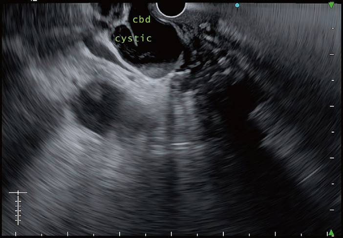
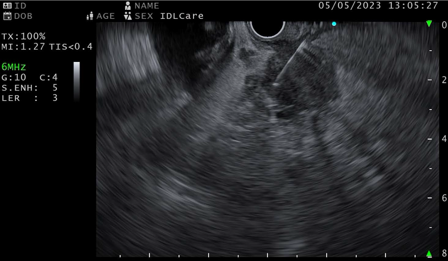
Evaluation of the needle visibility by size under EU-ME3
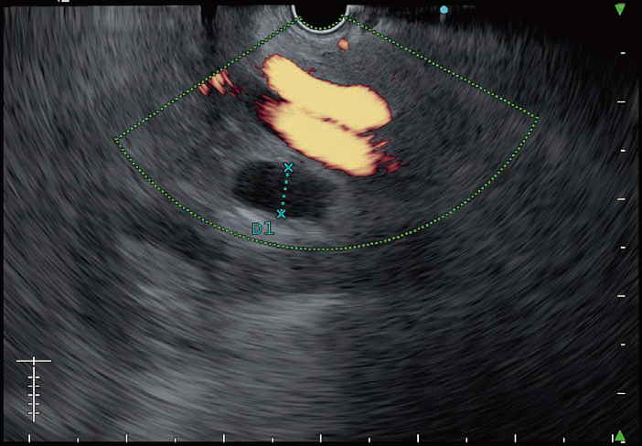
FNA/FNB needles under ultrasound image by EU-ME3 is clearly visible. We did several EUS-FNA/FNB procedures in pancreas and lymph nodes, and could identify the target lesion, needle and any structures in between more clearly compared to EU-ME2. Different sizes of needles were used for EUS-FNA/FNB and we can confirm that there was no noticeable deterioration of needle visibility even with smaller size of needles. The images on the right show Olympus FNB Needle, EZ Shot 3 Plus by different sizes (19G, 22G and 25G), under B-mode or THE-P mode of EU-ME3 during FNB procedures. Needle visibility under EU-ME3 is excellent and it can be seen end-to-end clearly even for 25G needle size. This end-to-end needle visibility helps to make EUS-FNA/FNB procedures safer with accurate targeting, supporting physicians to be more confident on making decisions. While using doppler functions, like color doppler, power doppler or H-flow, resolution of the image remains good without lag which possibly is because of high frame rate of EU-ME3.
After performing some EUS-FNA/FNB procedures using EU-ME3, we noted that its significant improvement supports not only better needle visualization but also better characterization of lesions thanks to higher resolution, clearer contrast and deeper penetration.
In addition, overall FNB work-flow has got much smoother and more responsive because of new user-friendly keyboard design with big touch panel and trackpad. Switching modes (e.g. from B-mode to doppler mode) is much easier without tapping the touch screen many times.
What are your final thoughts after using EU-ME3?
As a conclusion, I would like to say that the quality of image, as well as the ability of characterization of lesions, has significantly been improved from previous model.
In addition, introduction of brand-new feature in EU-ME3, typically Shear Wave Quantification (SWQ), will open newer dimensions like endo-hepatology, pancreatitis and pancreatic lesions to explore clinical solutions through EUS. New elastography in EU-ME3 has also become much useful with better stability thanks to i-ELST mode.
The high image quality makes interventional procedures safer with improved needle visibility, and more accurate targeting of the lesions. We expect those improvements on new generation EUS processor to contribute to better clinical outcome during our day-to-day procedures.
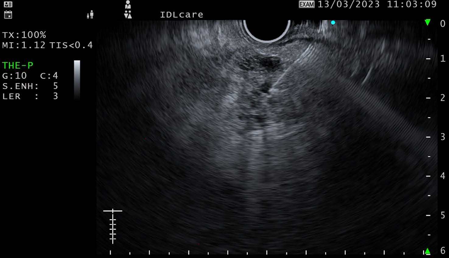
Dr. Thawee Ratanachu-ek How does EU-ME3 B-mode imaging, CHE, Elastography and SWQ improve EUS clinical practice?
Prof. Anthony Teoh
- Content Type