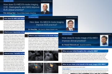
Prof. Anthony Teoh
Prince of Wales Hospital, Hong Kong
How does EU-ME3 add value to your clinical practice?
Endoscopic ultrasound plays a pivotal role in diagnosis and therapy of gastrointestinal conditions.
The procedure has evolved from being a diagnostic modality for fine needle biopsy, to a therapeutic intervention required for drainages of pancreatic fluid collections, biliary tract, gallbladder and gastrointestinal tract. The quality of imaging plays a key role in accurate diagnosis and therapeutic interventions. With superb B-mode and image enhancing features, the new EU-ME3 processor will add important values to my clinical practices.
What is your impression of B-mode image of EU-ME3?
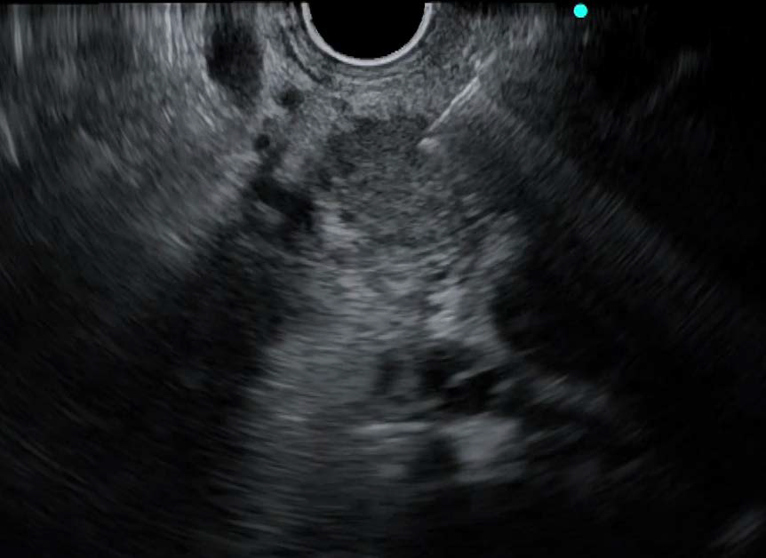
The quality of B-mode images is marked improved with higher image resolution and clarity. Furthermore, the new feature s-FOCUS reduces the change in resolution with distance from the transducer and eliminates the need to adjust focal zones during the procedure resulting in superior image qualities. I find it particularly useful in imaging deep seated lesions that are far away from the echoendoscope or inaccessible due to location of the lesion. The availability of tissue harmonic echo further comprehends the improvement in resolution, making images sharper for lesions such as tumour or pancreatic cyst.
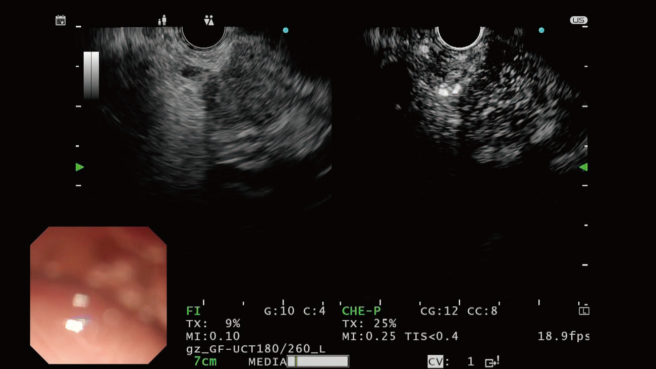
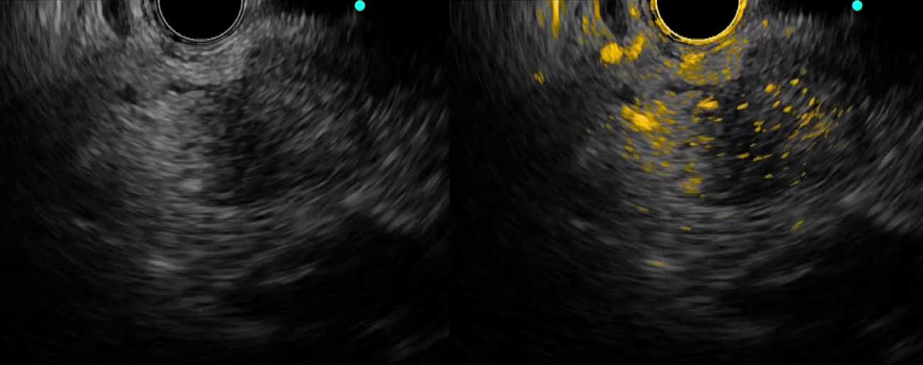
Clinical value / benefits of CHE
Contrast Harmonic Echo (CHE) allows the use of microbubbles to improve characterization of lesions as seen under EUS. When assessing the presence of mural nodules or suspected neuroendocrine tumours, the use of CHE can allow real time diagnosis of lesions without the need of biopsy. The new enhance CHE mode further improves resolution of the microvasculature seen in pancreatic tumours or cysts. We also use CHE to evaluate presence of viable tissue before and after EUS-guided radio-frequency ablation. The new overlay feature with colour coded contrast enhanced images allows easier visualization of the microbubbles in the vessels and differentiation from background black and white tissue. The time intensity curves (TIC) provides a method for quantifying of contrast intensity, much similar to contrast enhancements as seen in computed tomography or PET-CT.
This provides an objective method of measuring contrast enhancement intensity in malignancy and may provide a method of monitoring of the results of chemotherapy and anti-angiogenic treatments in unresectable patients with digestive cancers.
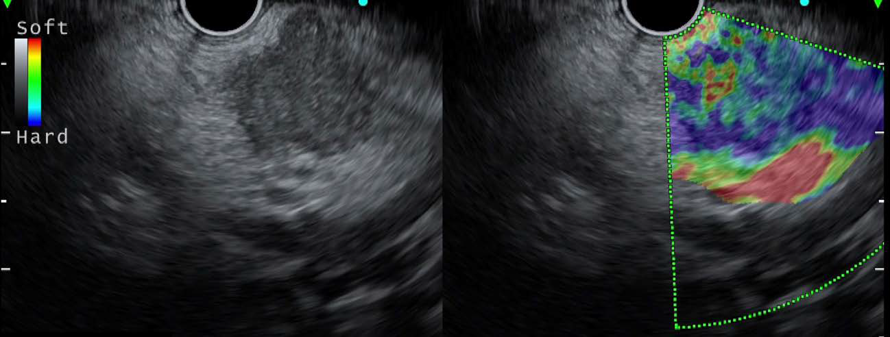
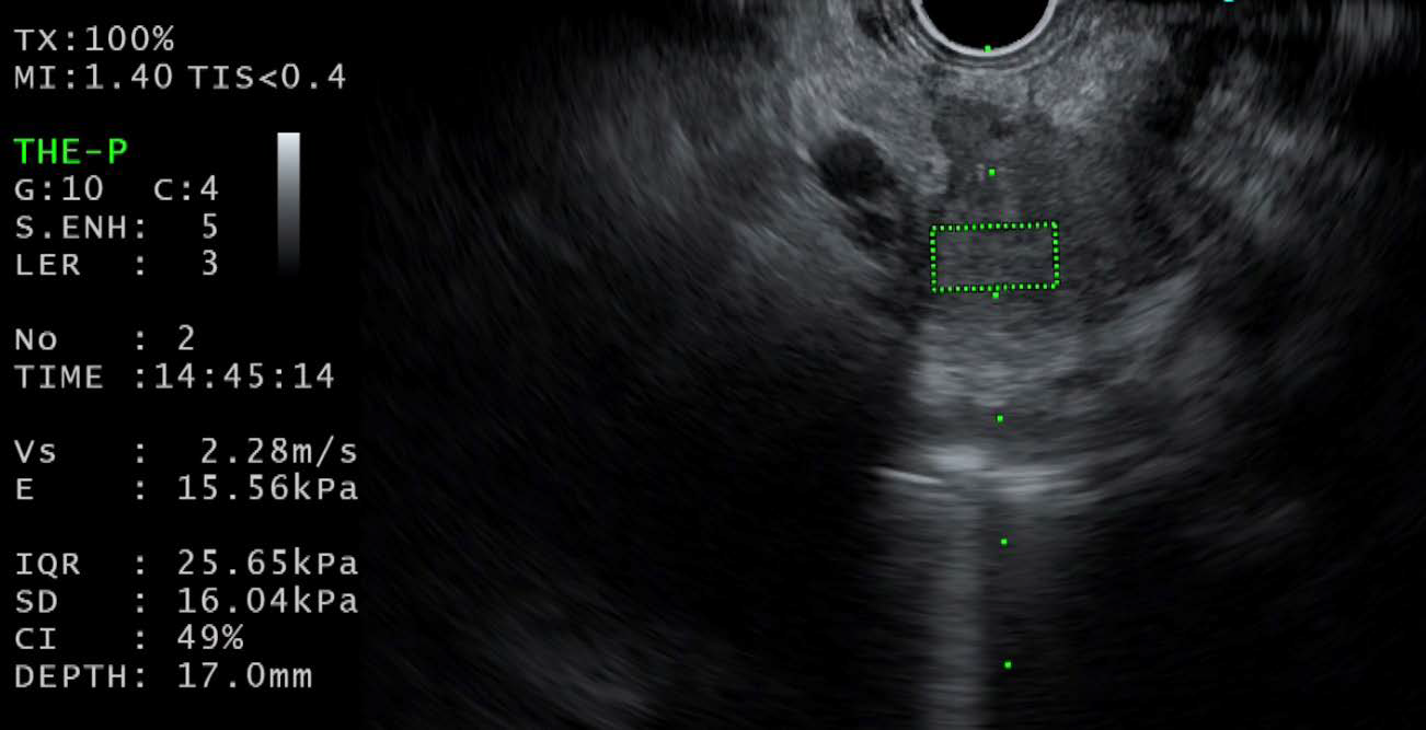
Clinical value / benefits of Elastography and SWQ
Elastography is a method to assess the hardness of tissue during EUS. However, the technique has been criticized previously for being too subjective and cannot provide quantitative measure of tissue hardness. The new feature of Shear Wave Quantification (SWQ) overcomes this weakness by providing an absolute value of tissue stiffness. This feature has great potential particular in the area of Endohepatology, where EUS can provide a quantification of liver stiffness, similar to the use of Fibroscan, providing another method to stratify patients with cirrhosis.
This feature allows a onestop option for endoscopic evaluation in patients with cirrhosis including endoscopic variceal surveillance, endoscopic measurement of liver stiffness and endoscopic measurement of portal pressure venous gradient.
Similarly, SWQ could be used for evaluation of pancreatic lesions with differentiation of pancreatic cancer from autoimmune pancreatitis or chronic pancreatitis.
- Content Type