Squamous Cell Carcinoma in Right Vocal Fold

Petr Lukes, M.D., Ph.D.
Department of Otorhinolaryngology, Head and Neck Surgery
First Faculty of Medicine
Charles University and University Hospital Motol
Endoscopic Finding;
Flexible video endoscopy with NBI- lesions visible on both vocal folds. Rough surface and reddening visible in white light image. Clearly visible margins of the lesion and pathological vascularization in NBI image.
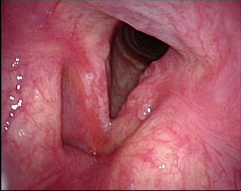
White Light Image
Equipment: OTV-S190, CLV-S190, ENF-VH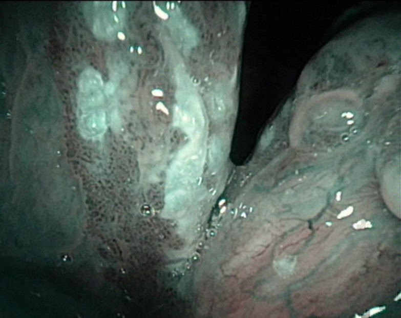
NBI Image
Pathological Finding;
Right vocal fold: severe dysplasia in the margins of the lesion with well-differentiated spinocellular cancer in the centre of the lesion
Left vocal fold: severe dysplasia
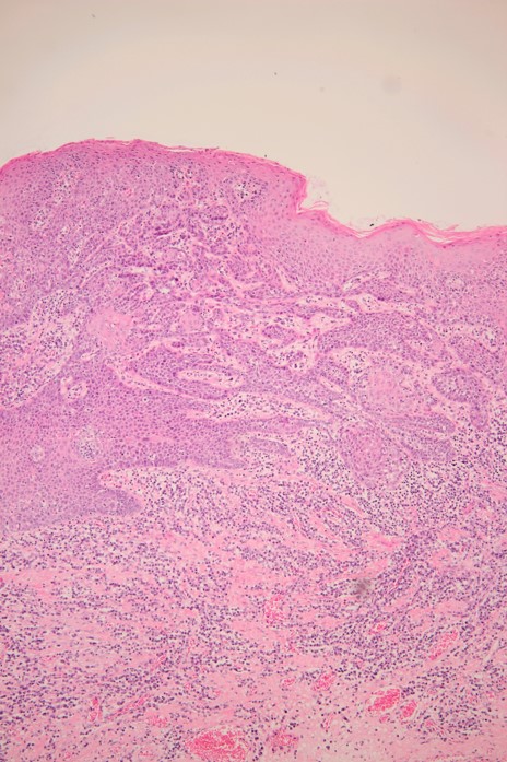
Pathological Image (macro)
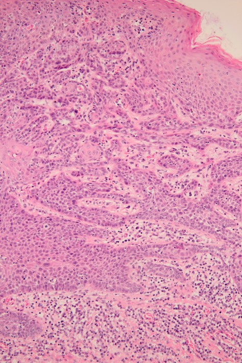
Pathological Image (micro)
- Content Type