NBI – The New Standard for Early Cancer Detection
NBI allows ENT specialists to diagnose and treat early laryngeal cancer even more precisely and reliably.
NBI is also a very accurate technology for follow-up procedures.
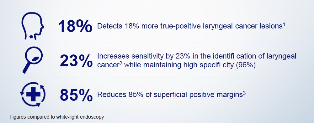
It is clinically proven1 that by using NBI, doctors are able to detect more suspicious lesions compared to traditional white light (WL), consequently leading to potentially reduced recurrence.4 In addition, NBI is easy to handle due to filter activation at the push of a button. And it’s cost-effective, too: All Olympus Medical imaging systems and videoscopes come with NBI as standard with no additional installations or drugs needed.
All this makes using NBI a safe and reliable way to improve clinical outcomes in patients with suspected cancer in the upper aerodigestive tract – and a unique tool for the patient pathway from diagnosis to follow-up in OR, outpatient clinics and doctors’ offices. It is suitable for numerous endoscopy procedures in ENT such as laryngoscopy, larynx, oral cavity, and sinus surgery as well as for procedures in otology.
NBI is a keystone in our daily practice: It is a reliable tool in the study of upper aerodigestive tract malignancies. (September 2018)
Prof. Giorgio Peretti
Professor of Otorhinolaryngology, University of Genova’s Medical and Postgraduate Schools,
Director of Otorhinolaryngology, Genova’s San Martino University Hospital
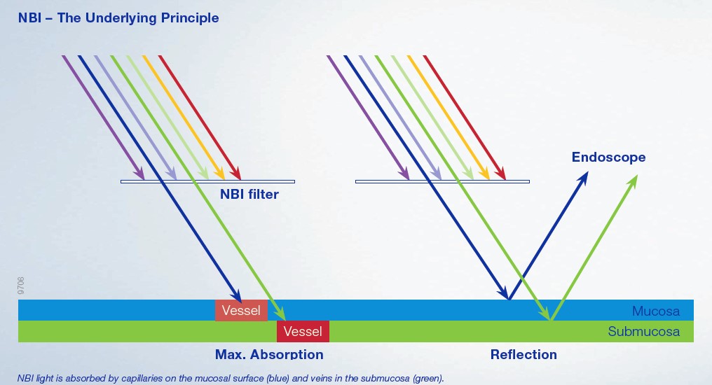
How NBI Works
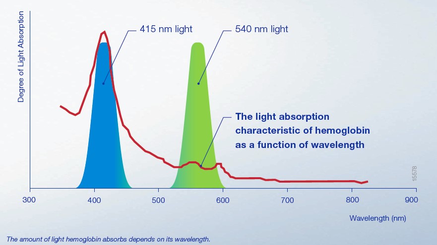
Normal white light (WL) contains all colors. When WL hits the surface of a tissue, all colors are absorbed. Thus the image remains with lack of contrast. With NBI this is different.
NBI uses only blue and green light. When blue and green light hit the surface of the tissue, it is highly absorbed by hemoglobin in the blood vessels. While the blue light is absorbed by the capillaries in the mucosa, the green light reaches deeper to the submucosal area, where it is reflected by the blood vessels.
This is why NBI creates a significantly higher contrast between blood vessels and the surrounding tissue than WL.
The NBI images therefore have more contrast than WL images.
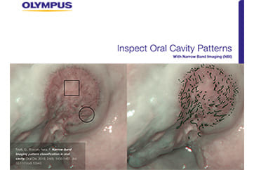
Since small tumors are often surrounded by many blood vessels, NBI helps to detect these tumors at an early stage and to analyze these areas accordingly. Thus, NBI supports the early and precise optical diagnosis of laryngeal cancer lesions, which as a result allows better treatment and more accurate follow-ups.
Numerous studies highlight the clinical value of NBI, especially with regard to the characterization of suspicious mucosal areas and the detection of cancerous lesions.
The Clinical Value of NBI for ENT
NBI is Clinically Proven to Diagnose More Laryngeal Cancer
Particularly when combined with a high resolution (e.g. HDTV), NBI can provide a more-detailed and higher-contrasted visualization of blood vessels than other endoscopic procedures.1
An example from daily clinical practice illustrates this: Before a patient’s vocal sound or vocal capabilities deteriorate, NBI can be routinely used to determine early changes in the vocal folds’ vessels, which can also be quantitively and qualitatively classified.
Numerous studies, including a growing number of randomized controlled trials (RCT) and metaanalyses, highlight the clinical value of NBI, especially with regard to the detection of cancer and through examination of suspicious mucosal areas. With high image quality and contrast in full HD or ultra high definition (4K), NBI can better display the edge of surgical margin and thus may reduce the number of biopsies.
NBI improves my diagnostic workup and optimizes endoscopy. (September 2018)
Prof. Dr. med. Christoph Arens
Director, University Hospital Magdeburg, Department of Otolaryngology
The Economical Value of NBI for ENT
How NBI Reduces Overall Costs
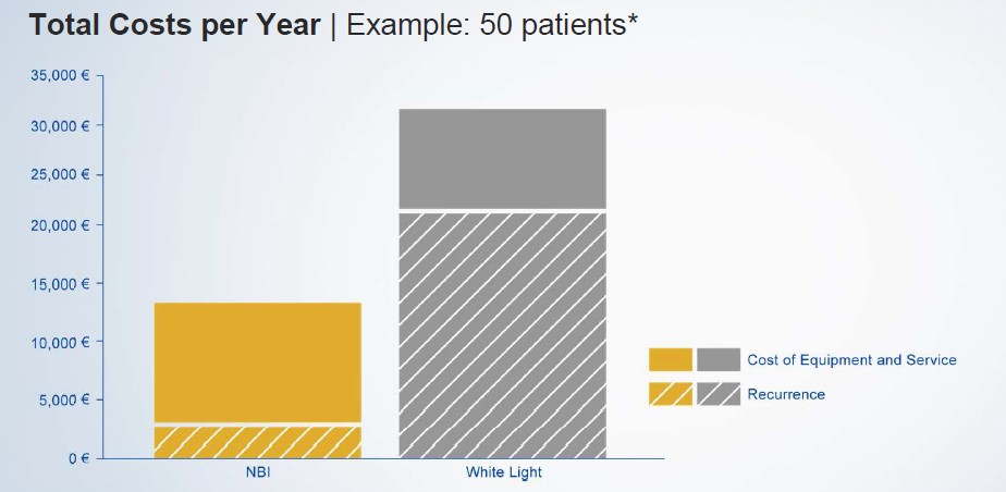
NBI offers superior efficacy in the early diagnosis of laryngeal cancer in pre-, intra- and postoperative stages compared to WL endoscopy. As tumors can be detected at an early stage using NBI, potential complications and the time a patient needs to spend in hospital for treatment, follow-up and treatment due to tumor recurrence is potentially less.4
NBI comes with Olympus Medical equipment at no additional cost for extra equipment, disposables or drugs. It is a key function of the Olympus endoscopy system and has been shown to provide clinical quality, efficiency gain and cost savings in other indications.5
1 Simo et al., European Laryngological Society: ELS recommendations for the follow-up of patients treated for laryngeal cancer. Eur Arch Otorhinolaryngol. 2014 Sep; 271(9): 2469-79.
2 Kraft et al., Value of narrow band imaging in the early diagnosis of laryngeal cancer. 2015 Wiley Periodicals, Inc. Head Neck 38: 15-20, 2016.
3 Garofolo et al., Intraoperative Narrow Band Imaging Better Delineates Superficial Resection Margins During Transoral Laser Microsurgery for Early Glottic Cancer. Ann Otol Rhinol Laryngol.2015 Apr; 124(4): 294-8.
4 Plaat et al.: Narrow-band imaging in transoral laser surgery for early glottic cancer in relation to clinical outcome. Head Neck. 2017 Jul; 39(7): 1343-1348. doi: 10.1002/hed.24773. Epub 2017 Mar 29.
5 Kang W et al.: Narrow band imaging-assisted transurethral resection reduces the recurrence risk of non-muscle invasive bladder cancer: A systematic review and meta-analysis. Oncotarget. 2017 Apr 4; 8(14): 23880-23890. doi: 10.18632/ oncotarget.13054.
In the following calculation, the Olympus video endoscopy system with NBI (“NBI”) will be compared to a conventional white-light endoscopy system without NBI (“White Light”).
Disclaimer: Costs and savings fi gures used in the model are for illustrative purposes only and the user’s attention is drawn to the fact that outputs from the model are subject to a) the assumptions described within the model and b) the data that the user selects or decides to input into the model.
* Plaat et al.: Narrow-band imaging in transoral laser surgery for early glottic cancer in relation to clinical outcome. Head Neck. 2017 Jul; 39(7): 1343-1348. doi: 10.1002/hed.24773. Epub 2017 Mar 29.
- Content Type