Superficial squamous cell carcinoma in the left pyriform sinus

Kenji Okami, MD, PhD
Professor,
Department of Otolaryngology,
Tokai University, Japan
Endoscopic Finding;
White light image demonstrate a small mucosal nodule in the left pyriform sinus. This is not the specific findings of malignancy. However, NBI image exhibited a well-demarcated brownish area with irregularly scattered vascular proliferation pattern on and around the nodule, which is the specific sign of the superficial cancer of the hypopharynx
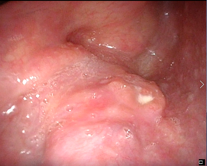
White Light Image
Equipment: OTV-S190, CLV-S190, ENF-VH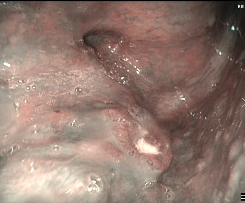
NBI Image
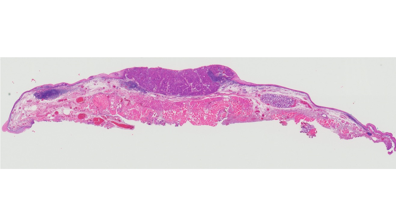
Pathological Finding;
Superficial squamous cell carcinoma with focally invading into the subepithelial layer. There was no muscular invasion. Tumor size was 12 mm in the greatest dimension and 2 mm in the thickness.
Both horizontal margin and vertical margin were negative.
Pathological Images(macro)
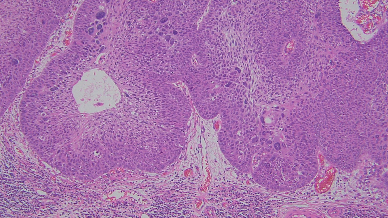
Pathological Images(micro)
Recurrent superficial cancer in the right piriform sinus after chemoradiotherapy
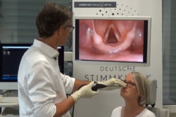
Squamous Cell Carcinoma in Right Vocal Fold
- Content Type