
Hospital: Thoraxklinik, University of Heidelberg, Germany
Scope: BF-1TH1100
Patient information:
63 years old male
Active smoker
Arterial blood pressure
Diabetes type II
Coronary arterty disease
COPD IIID
Medical history:
Hemopytsis (mild) since several weeks
Computertomographie showed emphesymatic changings, no nodule or enlarged nodes
1. WLI
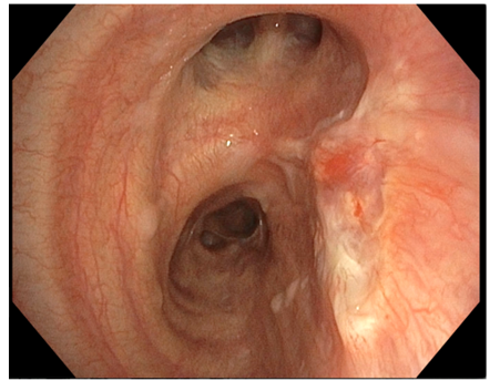
In white light an abnormal mucosa in front of the take off of the right upper lobe is visible.
2. TXI
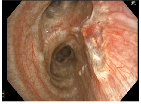
In TXI the borders are more clear and the pathological vessels at the corner become visible.
3. TXI
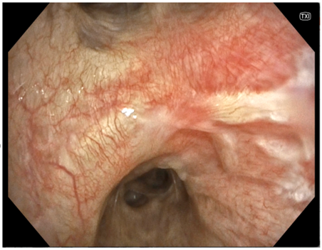
Here the border between the normal and abnormal mucosa is nicely seen.
Pathological Finding
The lesion was biopsied and a sqaumous cancer was detected. In the same session a radial EBUS was performed, showing a restriction of the cancer to the bronchial wall. Therefore, a Carcinoma in situ was diagnosed.
With the help of TXI the endoluminal boreders were visible as well as the malignant vasculary pattern.
Overall Comment
With the help of TXI the extent of the changings were better identifiable and the local staging with radial EBUS was more precisely.
With this information the tumor board was able to recommend a local endoscopic treatment (high dose radiotherapy) due to the comorbidities.
* Specifications, design and accessories are subject to change without any notice or obligation on the part of the manufacturer
- Keyword
- Content Type
