Case : Tumor in the trachea
Kei Morikawa, MD
Division of Respiratory and Infectious Diseases,
Department of Internal Medicine
St. Marianna University School of Medicine
Scope: BF-1TH1200
Location: Upper part of the trachea
Patient information: Male, 78 years old
Medical history: Intubation and tracheostomy were performed 2 years ago due to COVID-19 pneumonia. Chemotherapy was administered to treat mantle cell lymphoma 1 year ago. After complete remission of the lymphoma, PET-CT indicated a suspicion of a tumorous lesion on the right tracheal wall. He was referred to our institution for further examination. He is an ex-smoker who stopped smoking 3 years ago with a history of 27.5 pack-years.
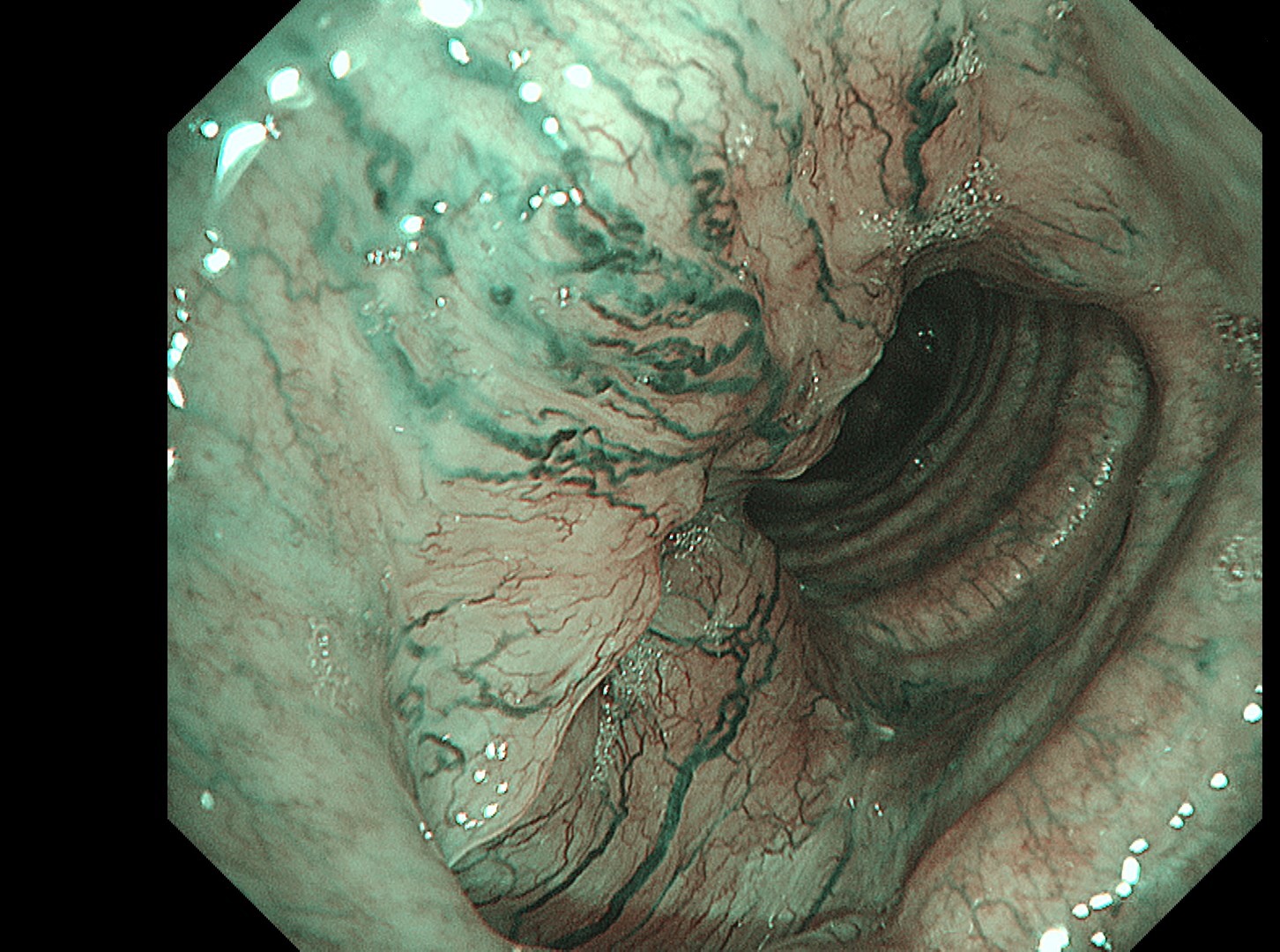
1-1 Upper part of the trachea (WLI)
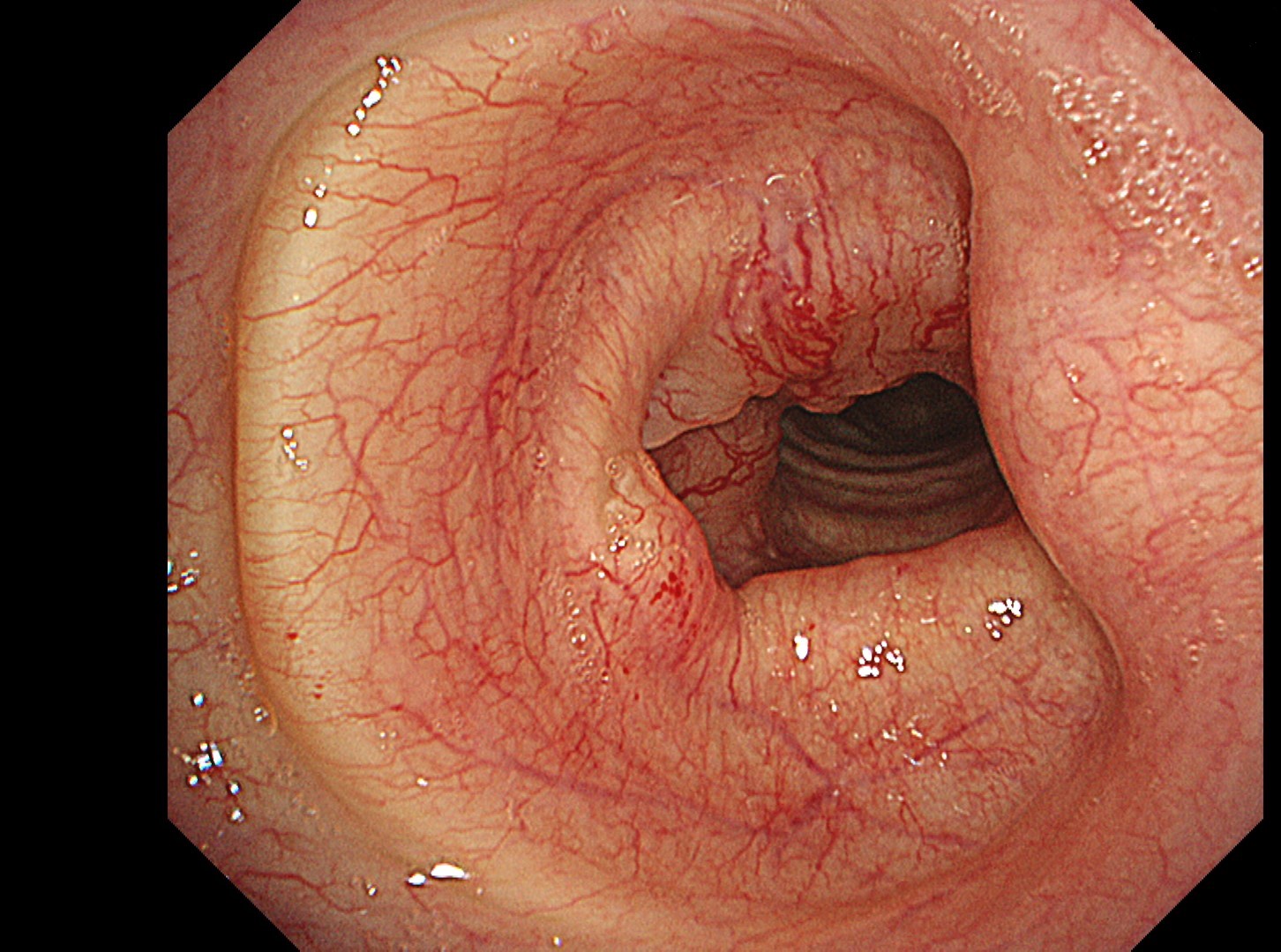
1-2 Upper part of the trachea (WLI)
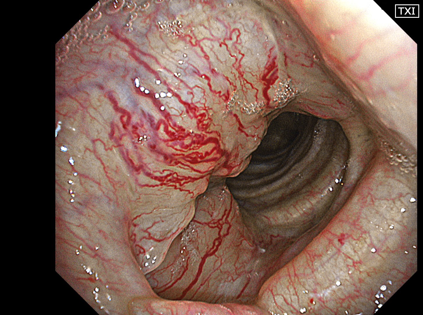
1-3 Upper part of the trachea (TXI1)
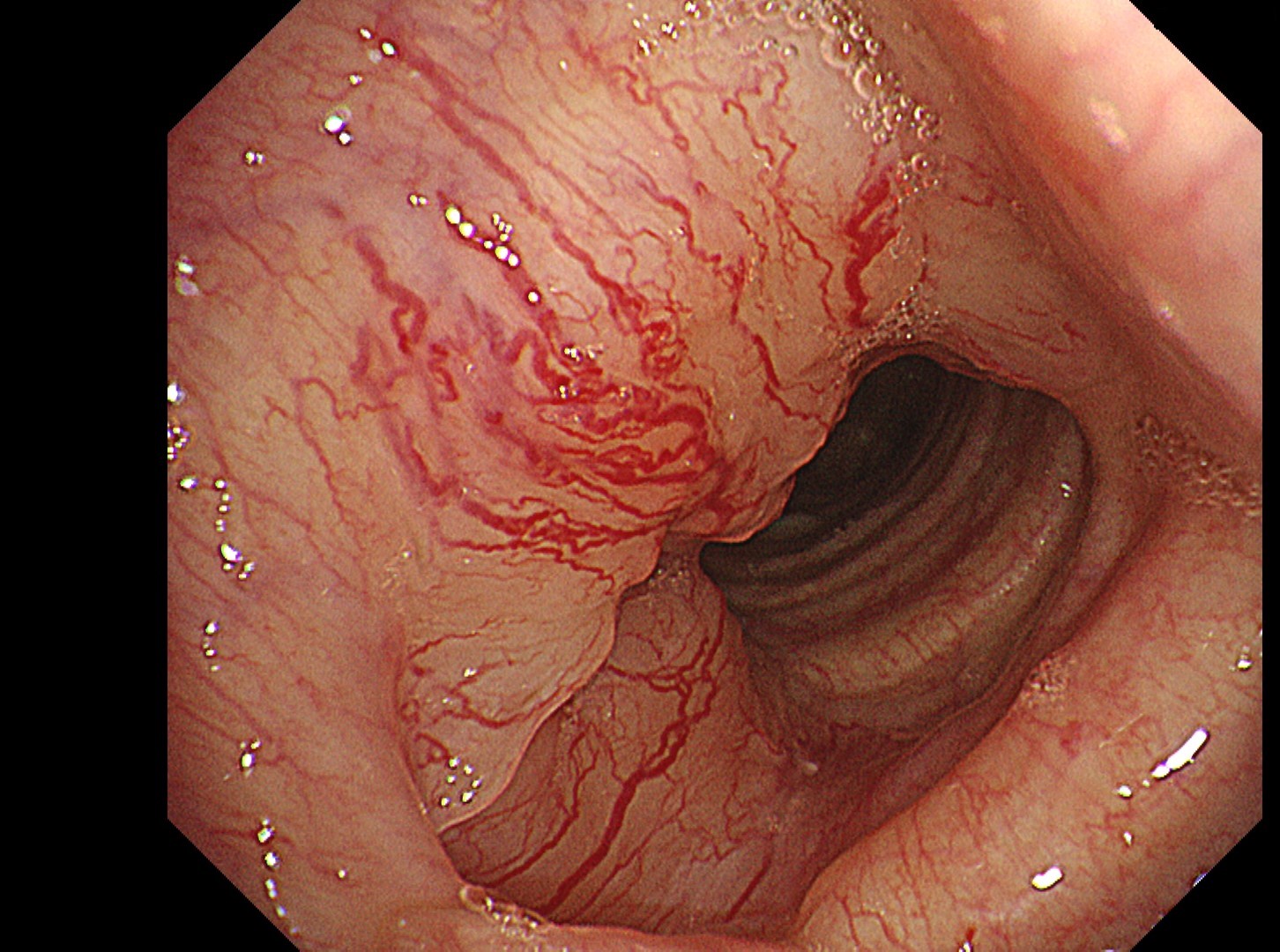
1-4 Upper part of the trachea (NBI)
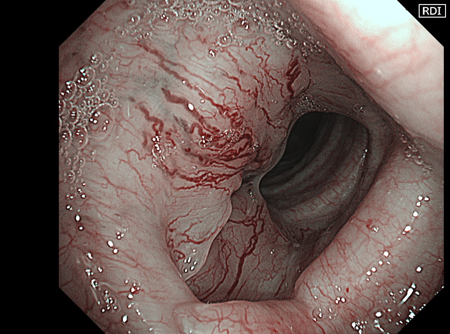
1-5 Upper part of the trachea (RDI2)
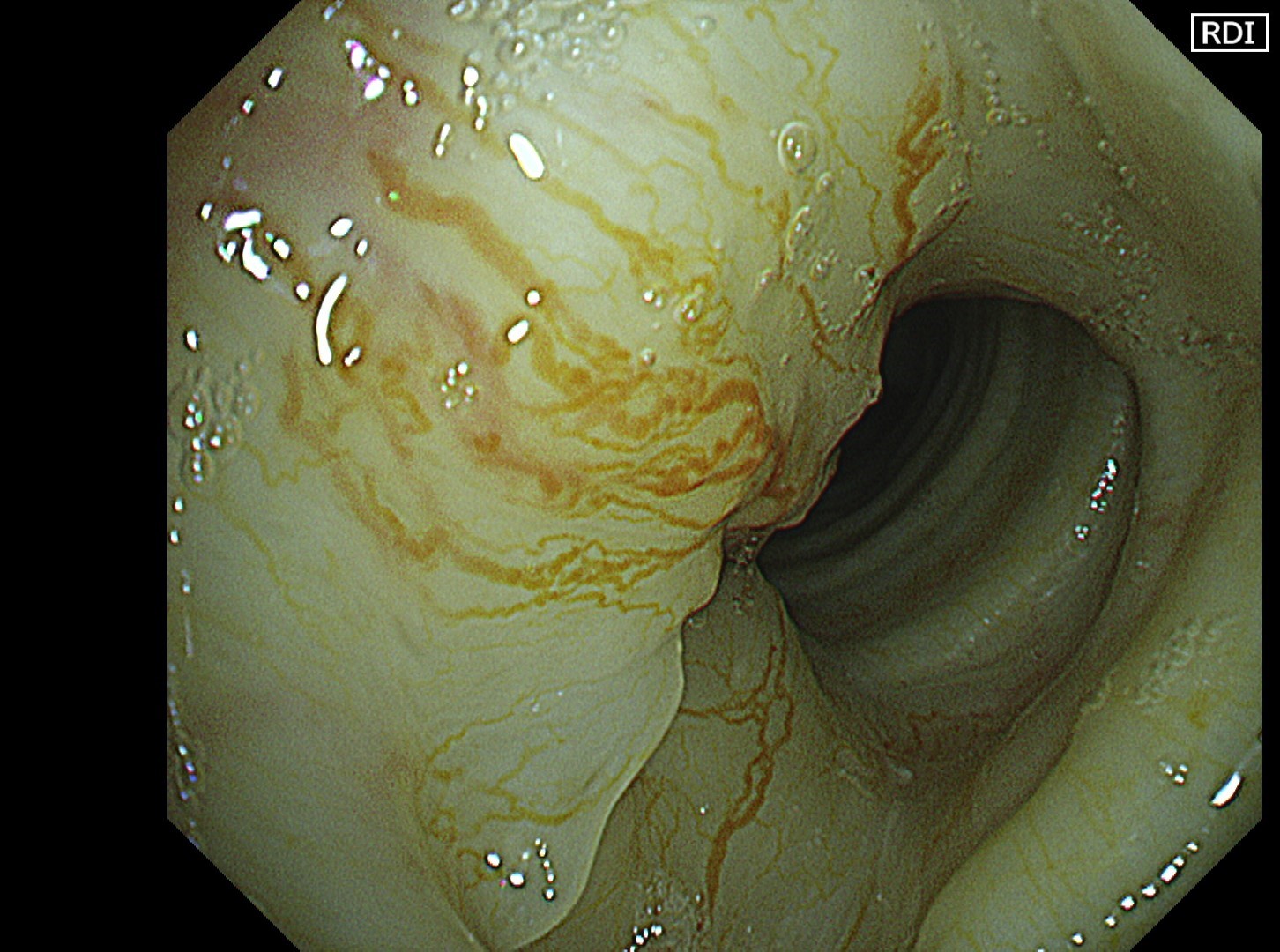
1-6 Upper part of the trachea (RDI3)
Case Video
The flat tumor growth in the upper part of the trachea shows a distribution pattern of subepithelial vessels suspected to be malignant and is carefully observed in detail in the NBI, TXI, and RDI modes. The lesion is subsequently biopsied while taking extra care not to cause bleeding.
Pathological Findings
- Figure A (HE staining): Tumor foci composed of proliferation of ductal epithelial cells and myoepithelial cells are observed. These feature a mesh-like structure including small cystic parts.
- Figure B (p63 immunostaining): p63-positive myoepithelial cells are observed at the periphery of the tumor foci.
- Final diagnosis: Adenoid cystic carcinoma


Overall Comment
In this case, abnormal subepithelial vessels that had increased irregularly were observed in detail in the TXI, NBI, and RDI modes. Since the lesion was located below the glottis, evaluation of the vessels in each mode was considered useful for careful selection of a biopsy site to prevent bleeding. The result suggests that the TXI and RDI3 settings, in particular, have the potential to be equivalent or superior to NBI for observation of vessels.
Co-editor:
Dr. Nobuyuki Ohike
Department of Pathology, St. Marianna University School of Medicine
- Content Type
