Case : Tumor in the right upper lobe

Fumihiro Asano, MD, PhD
Department of Pulmonary Medicine,
Gifu Prefectural General Medical Center
Scope: BF-1TH1200
Case:Tumor in the right upper lobe
Location: Right upper lobe bronchus
Patient information: Male, 70 years old
Medical history:Chest CT during his health checkup revealed a mass shadow in the right pulmonary hilum, and he was referred to our institution.
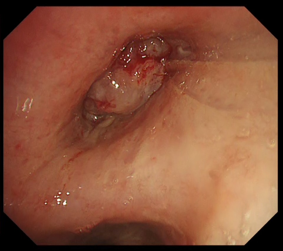
1-1 Tumor in the right intermediate trunk (WLI)
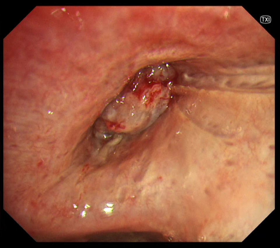
1-2 Tumor in the right intermediate trunk (TXI)
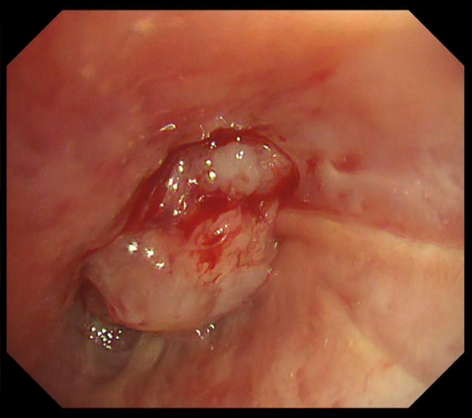
1-3 Close-up of the tumor in the right upper lobe (WLI)
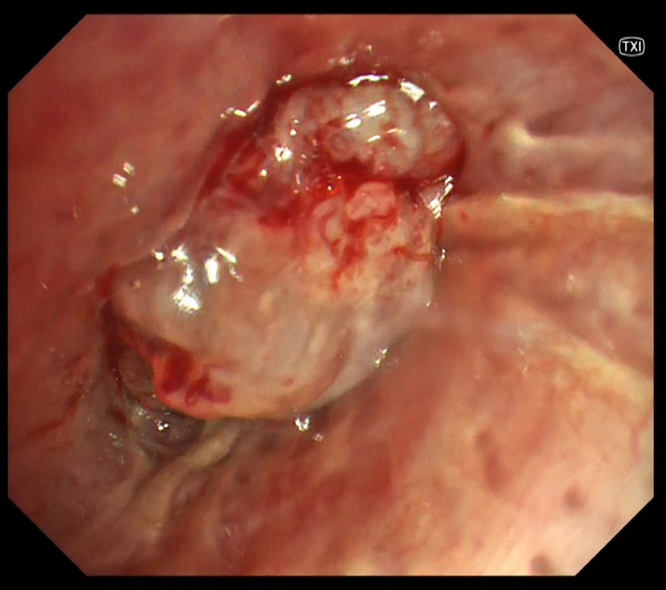
1-4 Close-up of the tumor in the right upper lobe (TXI)
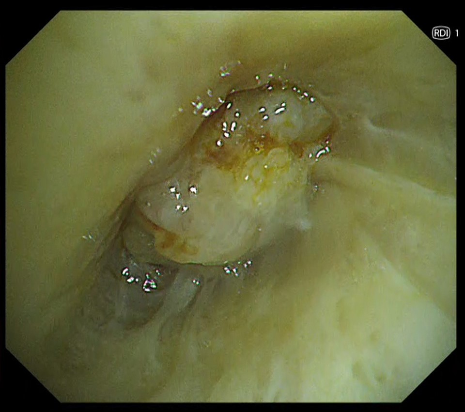
1-5 Close-up of the tumor in the right upper lobe (RDI)
Case Video
Pathological Findings
- Lung squamous cell carcinoma (diagnosed with TBB of the right upper lobe tumor and EBUS-TBNA of metastasized mediastinal lymph node)
- Proliferation of lung squamous cell carcinoma with a tendency to keratinization is observed.
Overall Comment
This was a case of squamous cell carcinoma of the lung (cT1N2M0 stage, IIIA). Bronchoscopy showed that the advanced peripheral squamous cell carcinoma partially progressed towards the central bronchi in a polypoid fashion. The convergence of longitudinal folds suggested that the upper right lobe bronchus was narrowed in a pointed fashion and the lesion extended around the bronchus. In TXI, structures such as longitudinal folds and fine vascular networks on the mucosa were more enhanced and could be observed more clearly than with white light.
- Content Type
