Hospital: Thoraxklinik, University of Heidelberg, Germany
Scope: BF-1TH190
Patient information:
58 years, male
Ex smoker (45 packyears)
Coronary artery disease
Medical history:
Patients reported on chronic coughing,
Lowdose computertomograhy showed no lung abnormalities
Lung function: FEV1 63%
1. WLI
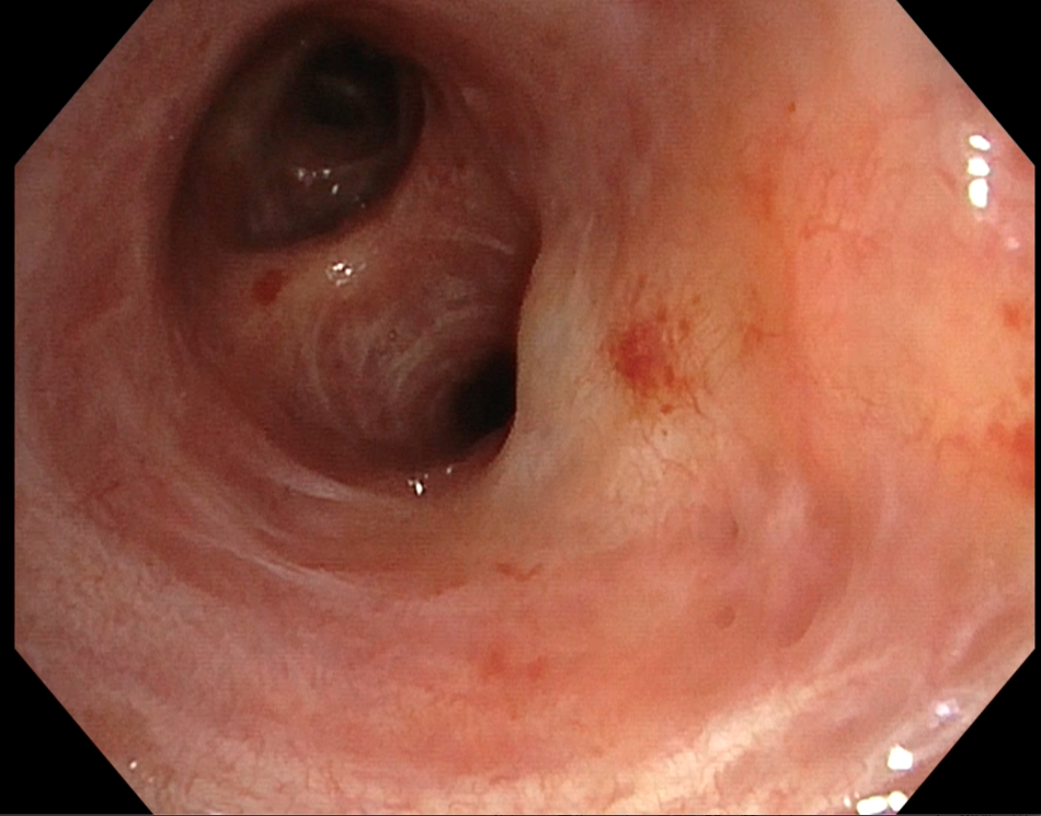
In the white light mode vasculary engorgement was visible.
2. TXI
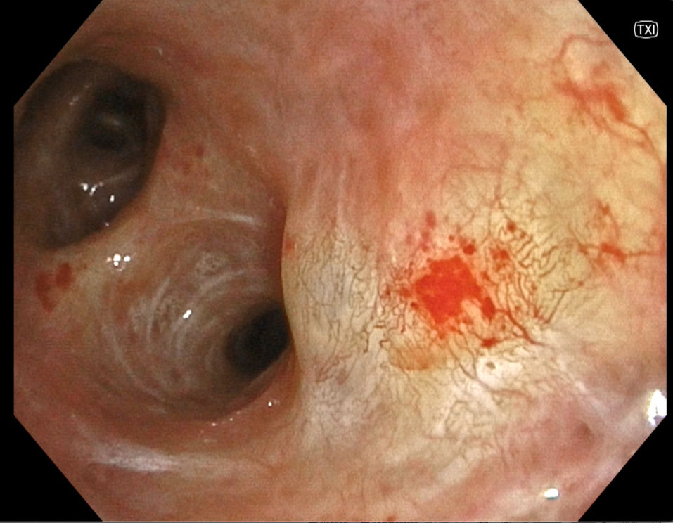
In TXI the vaculary pattern was seen more clear, vasculary slings as well as dotted vessel are visible.
3. RDI Mode 2
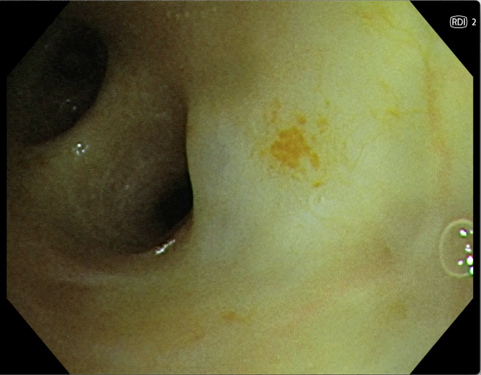
In RDI 2 especially the dotted vessels are highlighted.
4. RDI Mode 3
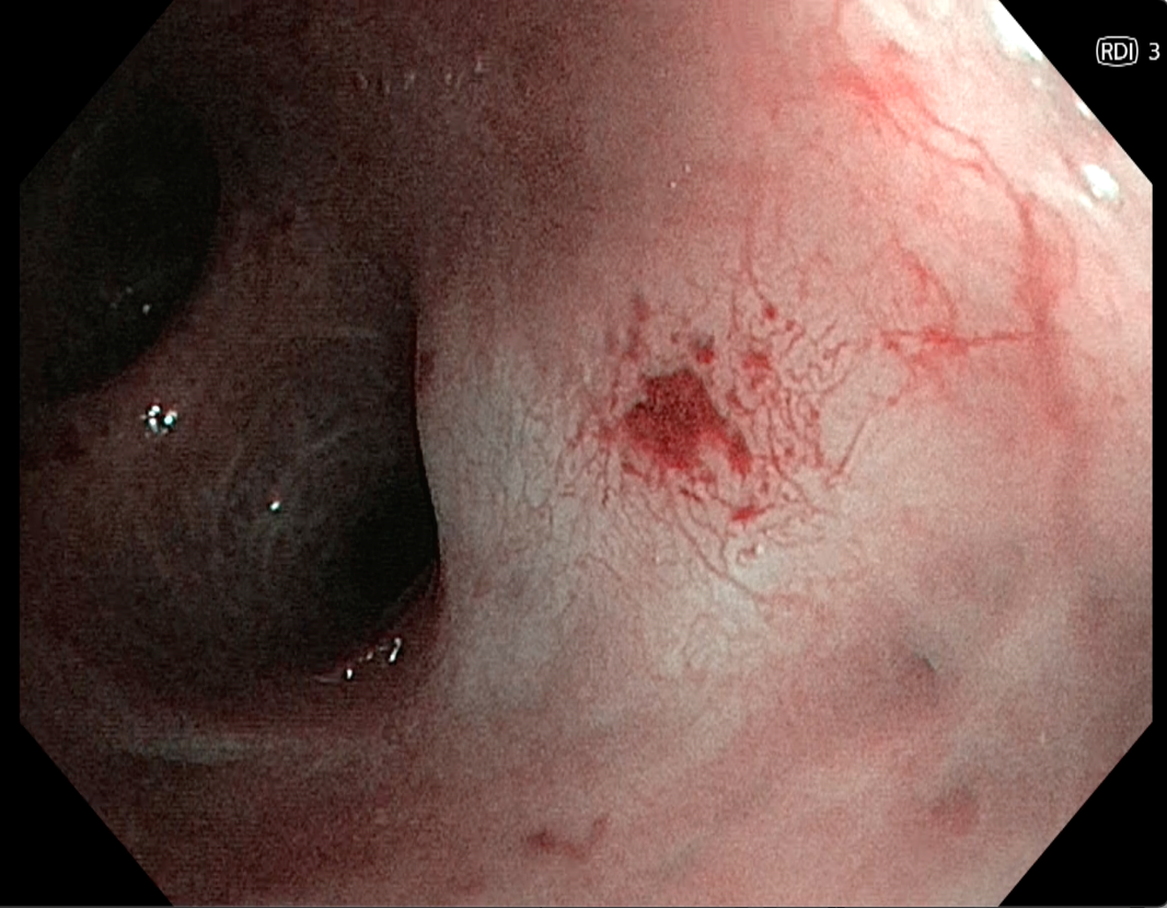
RDI 3 offers an alternative mode showing comparable patterns like TXI in this case.
Pathological Finding
Vasculary changes have been seen at the middle lobe entrance.
With the help of TXI and RDI 3 the vasculary pattern was seen nicely and targeted biopsies have been taken, showing a moderate dysplasia.
Overall Comment
With the help of the new features the pathological finding was better visible and it was possible to distinguish normal and abnormal areas.
The positioning of the forceps for the biopsy was more precisely.
* Specifications, design and accessories are subject to change without any notice or obligation on the part of the manufacturer
- Keyword
- Content Type
