Case : Snare resection of tumor in the proper segment of the left upper lobe

Li Shiyue Professor Guidance Expert
The First Affiliated Hospital of Guangzhou Medical University

Tang Chunli Professor Operation Expert
The First Affiliated Hospital of Guangzhou Medical University
Scope: BF-Q290
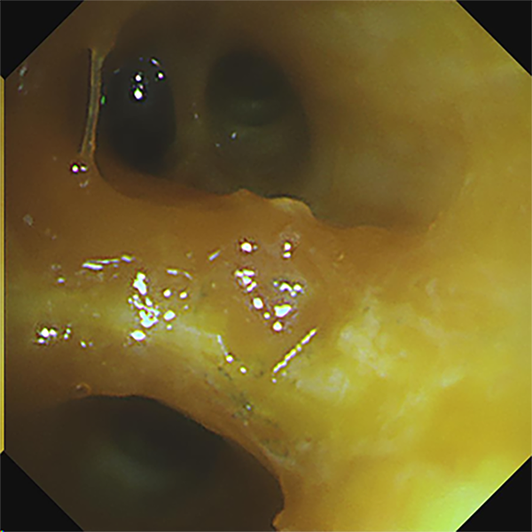
Location: Left upper lobe
Patient information: Male, 18 years old
Medical history: Intermittent coughing for two years, hemoptysis for one week, no fever, chest pain, or dyspnea. Chest CT indicates a neoplasm in the left upper lobe.
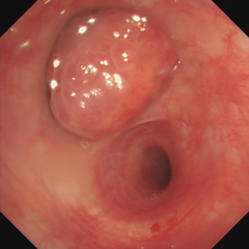
1. Left upper lobe (WLI)
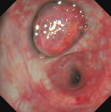
2. Left upper lobe (TXI)
3. Left upper lobe (RDI)
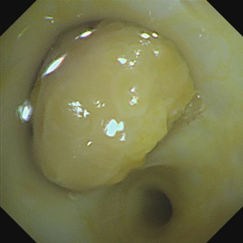
4. After snare resection of the tumor in the proper segment of the left upper lobe (WLI)
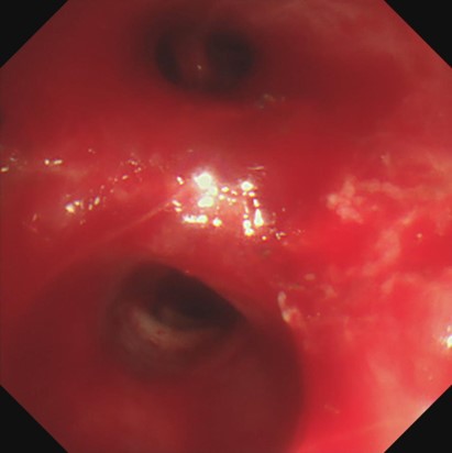
5. After snare resection of the tumor in the proper segment of the left upper lobe (RDI)
Case Video
The bleeding site can be clearly displayed in RDI mode
Hemostasis of active bleeding
Pathological diagnosis
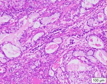
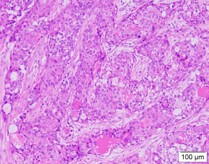
- Two pieces of gray-white tissues (1.2cm×0.8cm×0.5cm and 1.5cm×1.0cm×0.5cm in size) in the proper segment of the upper lobe of left lung was seen with the naked eye (full covered)
- Endoscopic findings: The tumor cells in the tissues (of the proper segment of the upper lobe of left lung) examined were composed of nonkeratinized squamous cells with the shape of sheet and polygon, intermediate cells, and mucus-secreting columnar cells. Most of the intermediate cells had round or oval nuclei with nucleoli, and a small number were columnar cells that secrete mucus, forming small glands, tubules or cysts. Nuclear division was not obvious and no necrosis was observed.
- Molecular pathology results: MAML2(+)
- Immunohistochemistry results: TTF1(-), NapsinA(-), P40(partially+), SMARCA4(+), ALK-P(D5F3)(-), ALK-P(Neg)(-), Ki67(hot-spot 15%+),PD-L1(E1L3N)(-),PD-L1(Neg)(-),CK5/6(+),CK7(+)
- Special staining results: Mucicarmine (+)
- Pathological diagnosis: Tissue (of the proper segment of the upper lobe of left lung) changes in line with bronchial low-grade mucinous epidermoid carcinoma
Overall Comment
Compared with the WLI mode of the bronchoscope, the TXI mode can display deeper vascular structures, which help determine the treatment of lesions. If bleeding occurs during treatment or biopsy, the RDI mode can help avoid the impact of blood on the bronchoscope camera. Especially for beginners, it can reduce the effect of blood contamination on the camera’s field of view, and help them easily see the bleeding site and accurately implement local hemostasis at the same time.
- Content Type
