Case : Right lung tumor

Fumihiro Asano, MD, PhD
Department of Pulmonary Medicine,
Gifu Prefectural General Medical Center
Scope: BF-1TH1200
Case:Right lung tumor
Location: Between the right intermediate trunk and the right upper lobe bronchus
Patient information: Female, 70 years old
Medical history:While she was receiving treatment for emphysema at a local clinic, chest X-ray revealed a right lung mass and pleural effusion. She was then referred to our institution.
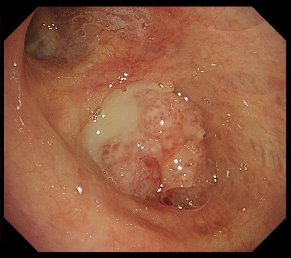
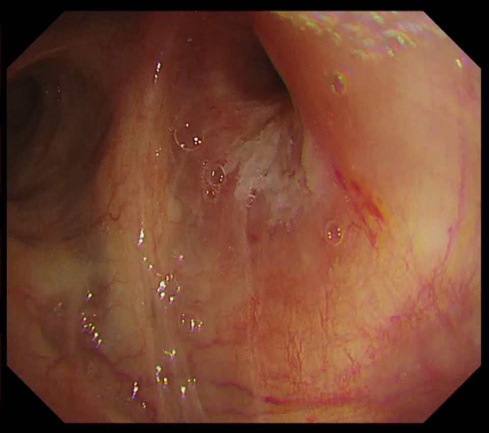
1-1 Tumor in the right intermediate trunk (WLI)
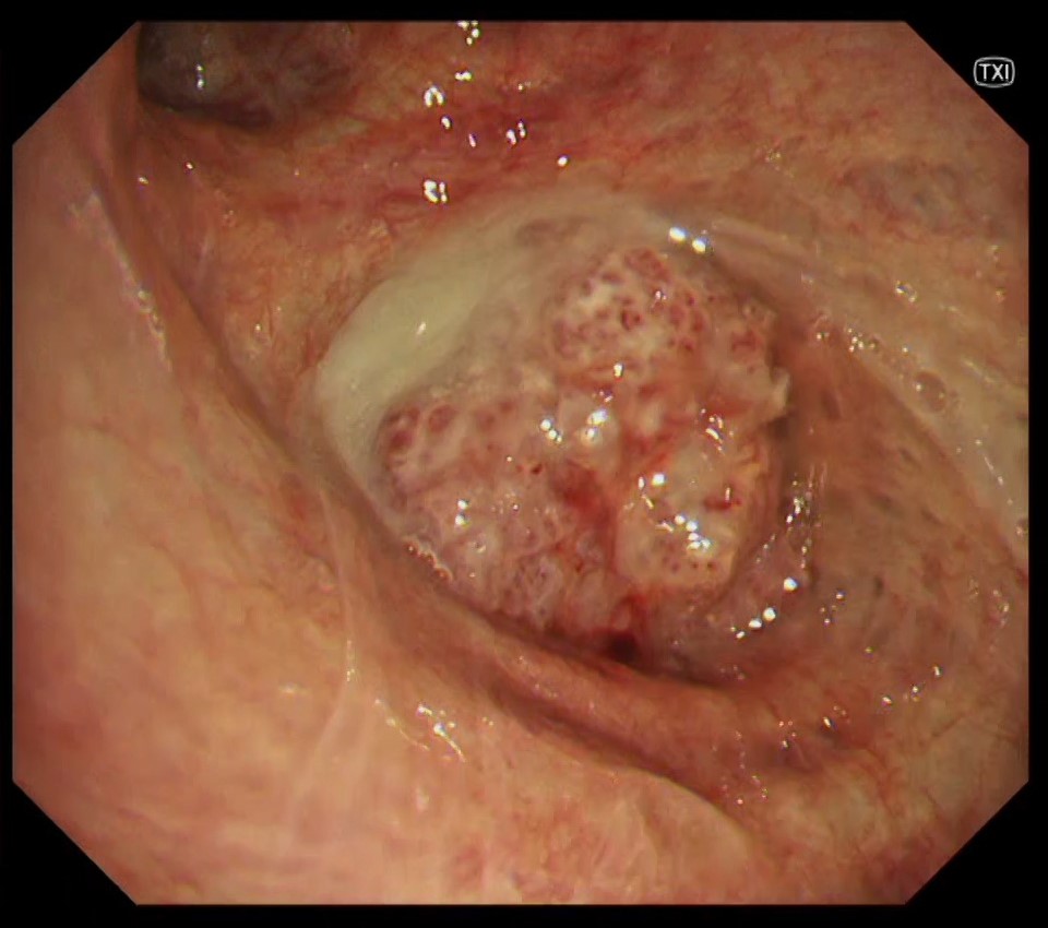
1-2 Tumor in the right intermediate trunk (TXI)
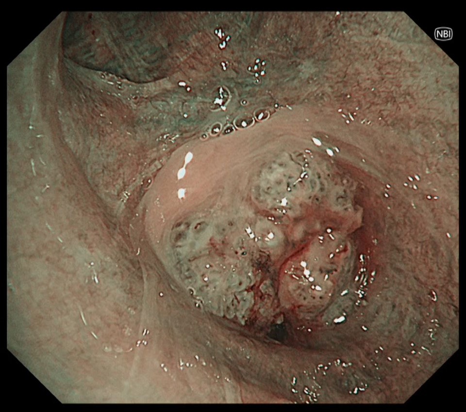
1-3 Tumor in the right intermediate trunk (NBI)
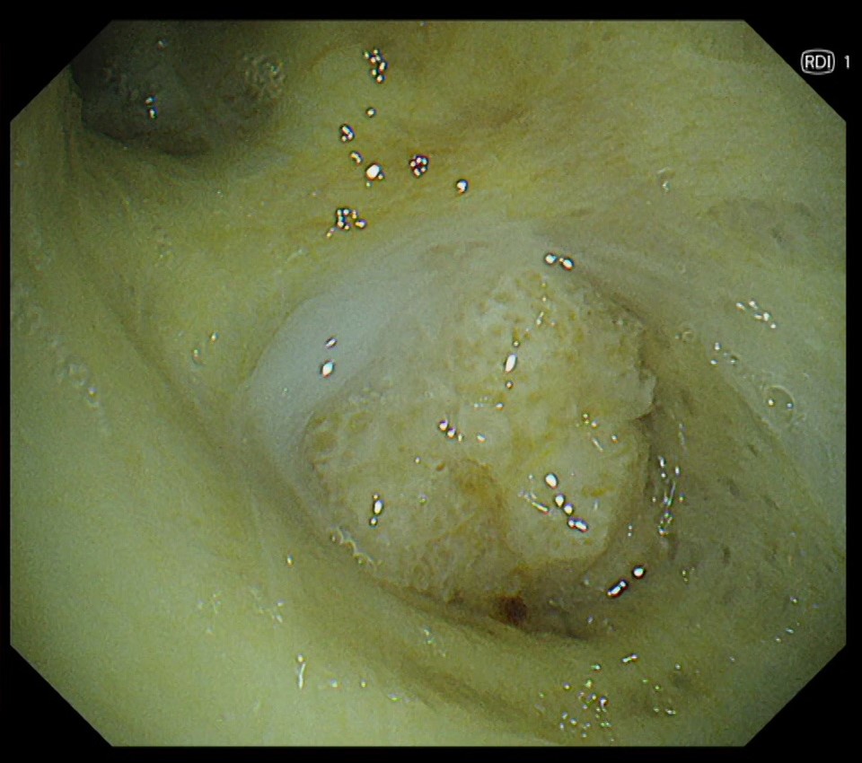
1-4 Tumor in the right intermediate trunk (RDI)
1-5 Lesion in the right upper lobe bronchus (WLI)
1-6 Lesion in the right upper lobe bronchus (TXI)

Case Video
Pathological Findings
- The tumor located in the intermediate trunk was biopsied endoscopically and diagnosed as squamous cell carcinoma with keratinization.
- The tumor cells tested positive for p40 but negative for TTF-1.
Overall Comment
This case is a lung squamous cell carcinoma originating from beyond the intermediate bronchus, with suspected infiltration into the upper lobe bronchus wall from lymph node metastasis in the hilum. TXI has the advantage of observing not only abnormal findings such as red dots and bridging fold, but also the intricate vascular network of normal areas in greater detail compared to WLI. This makes it easier to comprehend the extent of the lesion’s infiltration.
- Content Type
