
Department of Respiratory and Critical Care Medicine Director,

Department of Respiratory and Critical Care Medicine
Scope: BF-1TH1100
Patient information: Female, 61 years old
Medical history: Referral from our outpatient clinic to follow up an atelectasis of the left upper lobe caused by a central tumor mass.
1. WLI
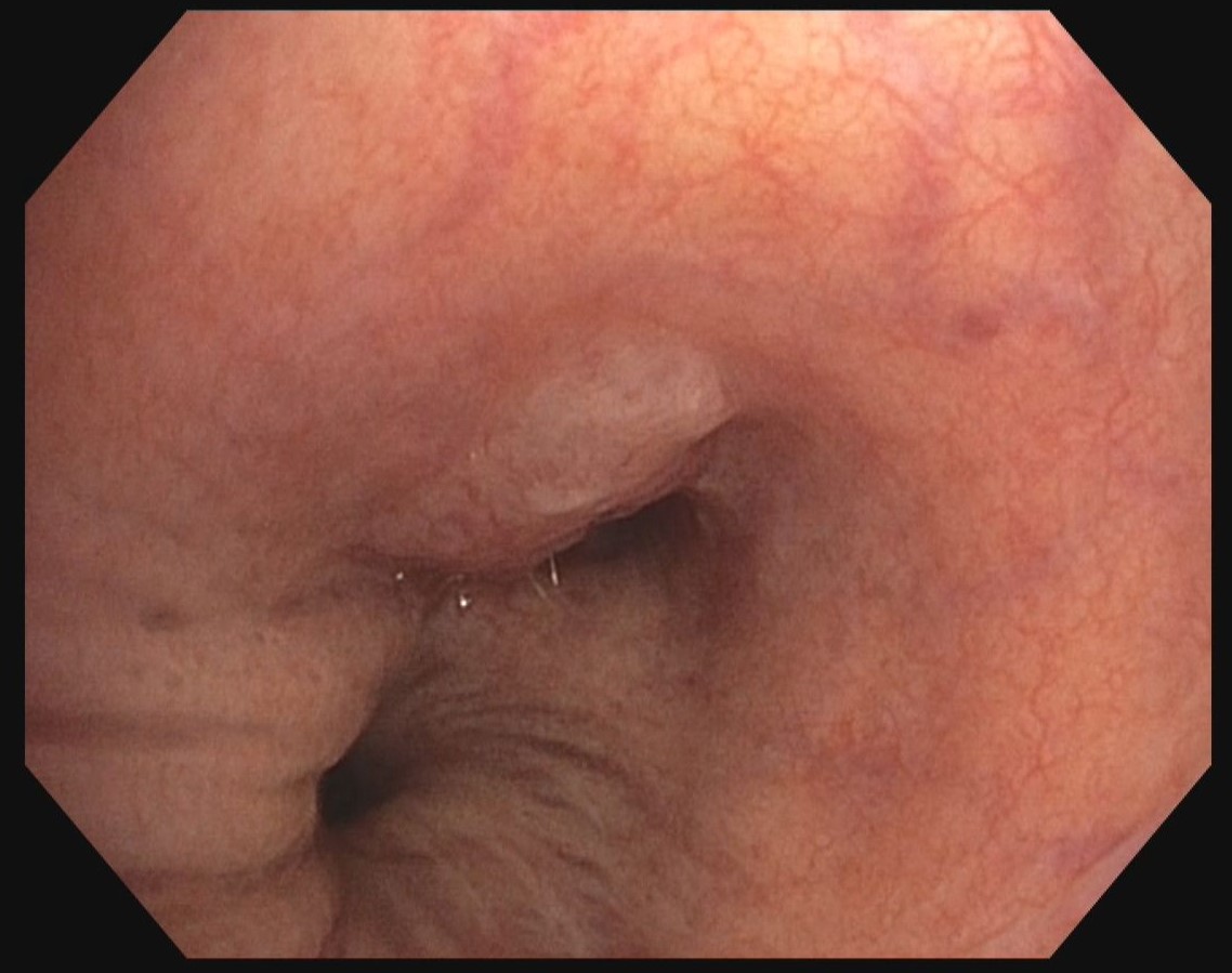
Mucosal tumor mass in the transition area between the distal left main bronchus and the left upper lobe stem bronchus.
2. TXI
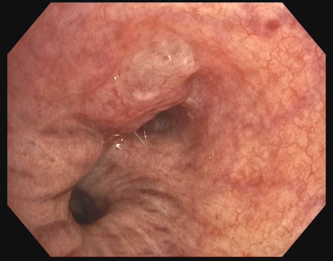
Better visibility of the mucosal tumor mass in the transition area between the left main bronchus and the left upper lobe stem bronchus. Even vascularisation of the tumor mass is highlighted.
3. NBI
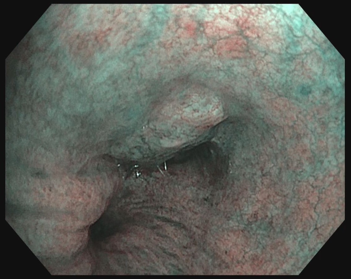
Improved visibility of the blood vessels and the mucosal tumor mass.
4. RDI
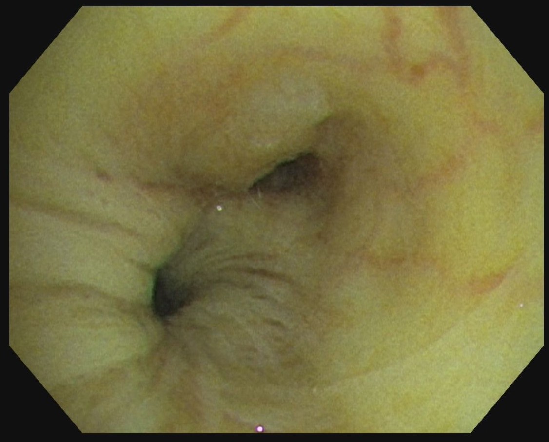
A better visibility of the large vessels facilitates performing safe biopsies of the tumor tissue.
Case video
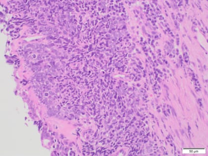
Pathological Findings
Lung biopsy tissue with formations of small cell lung cancer (NEC 3), bronchial mucosa with squamous cell metaplasia.
Overall Comment
Examination of the transition area between the lateral wall of the distal left main bronchus and the left upper lobe revealed a plain submucosal foreign tissue infiltration extending to a convex stenosis of the lumen of left upper lobe stem bronchus. This results in a subtotal occlusion of the left upper lobe. Especially TXI mode highlights visibilty of mucosal foreign tissue mass in this case.
* Specifications, design and accessories are subject to change without any notice or obligation on the part of the manufacturer
- Keyword
- Content Type
