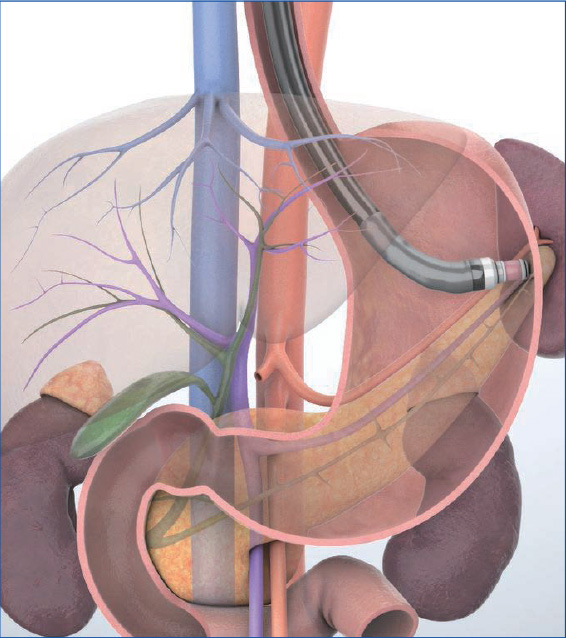
Imaging Techniques Transgastric approach
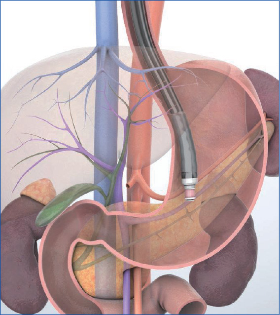
Straighten the scope in the stomach to observe sectional images perpendicular to the scope when viewed from the foot of the patient. To ‘The common method is to first advance the scope to the gastric body or antrum and scan while pulling the scope with the balloon inflated. Another method is to perform continuous scanning while in the “short-scope” position during transduodenal maneuvering. Either way, when the pancreas is visualized, position the pancreas in the six o’clock position on the screen and observe the body and tail of the pancreas and its vicinity while using anatomical features in the surrounding area as landmarks.
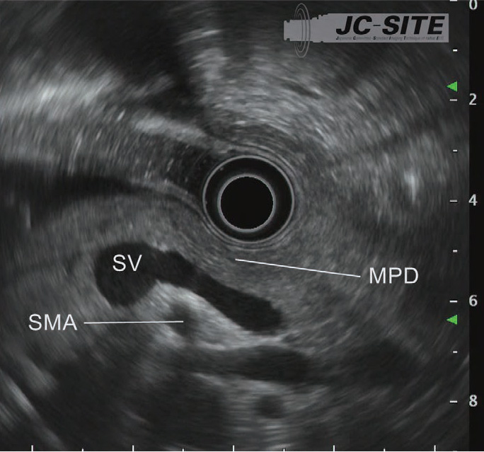
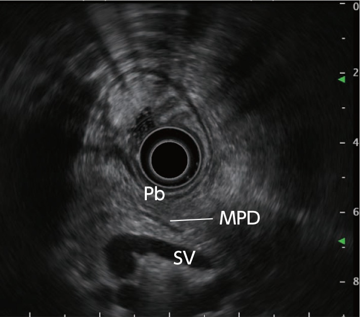
STEP 1 | Body of the pancreas
Slowly pull the scope from the gastric body or antrum to straighten the scope. Then use the splenic vein as a landmark to observe the pancreatic body.
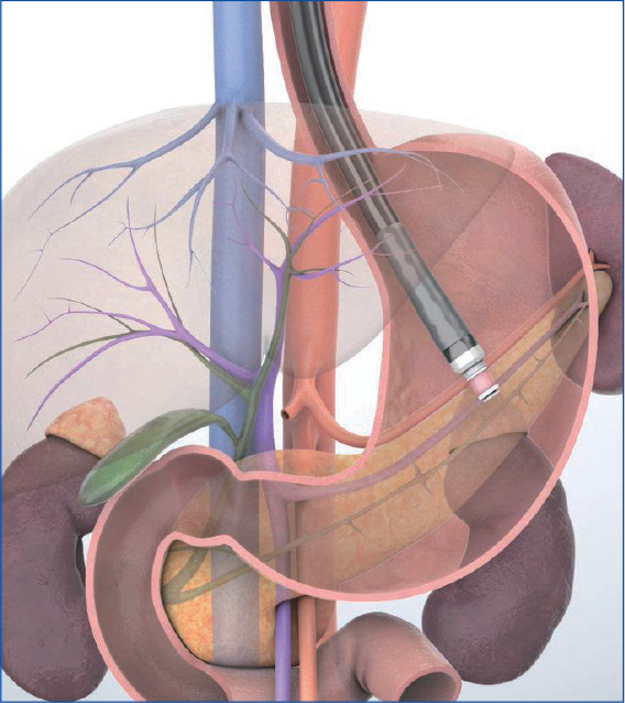
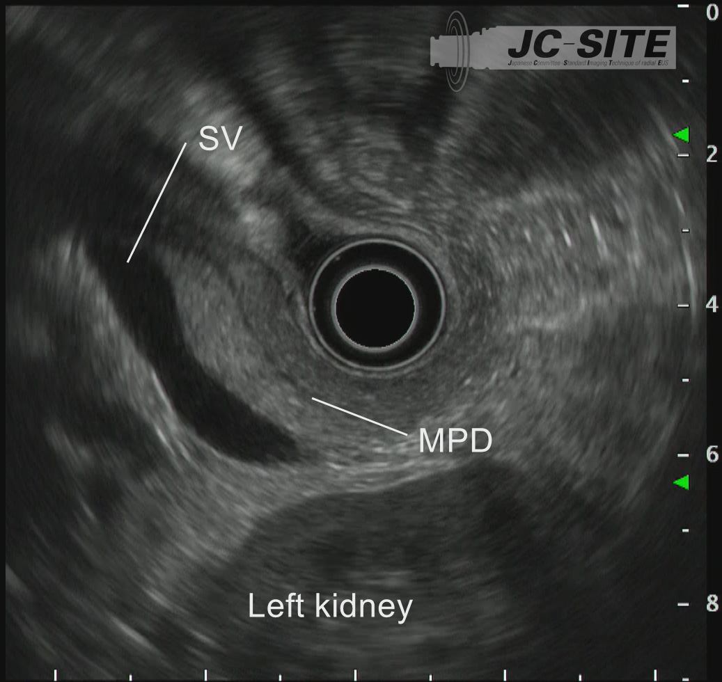
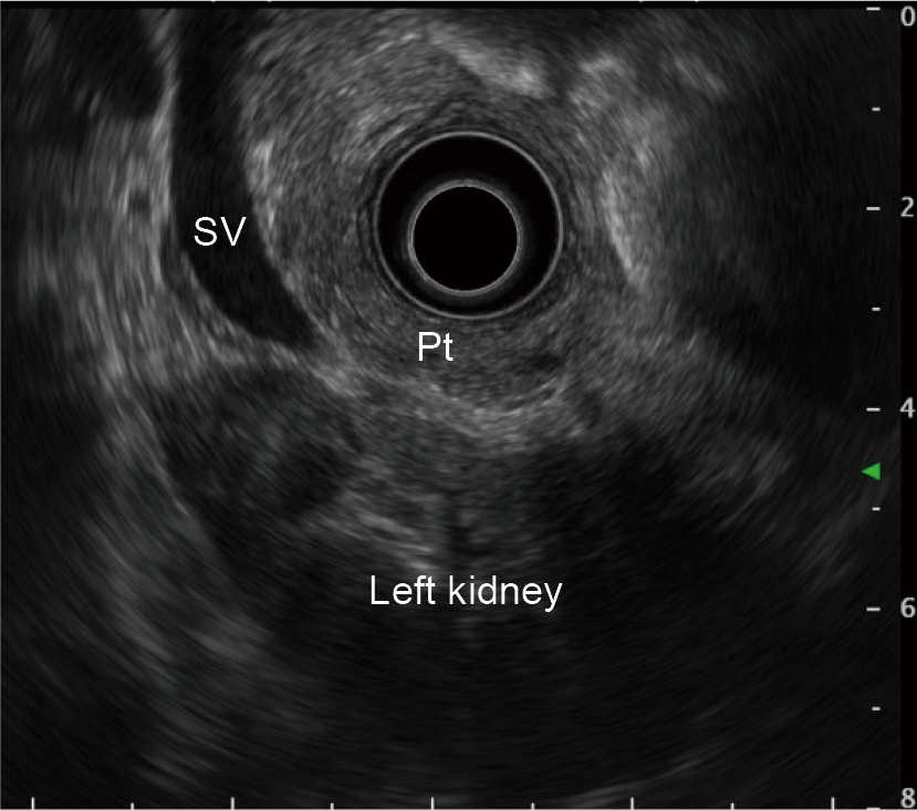
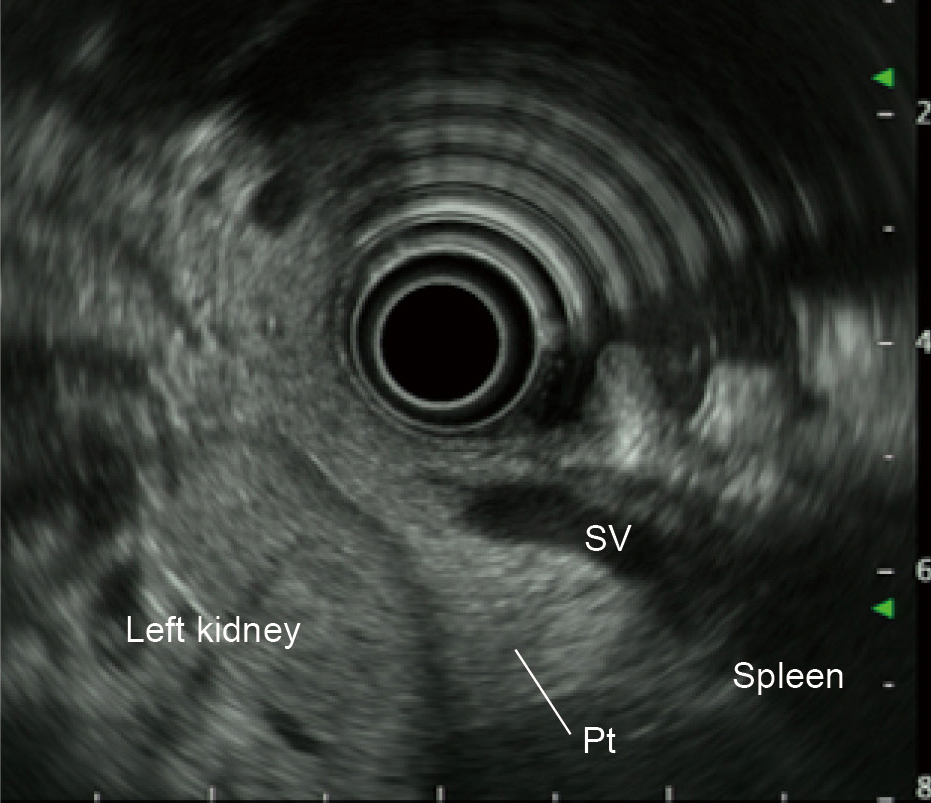
STEP 2 | Tail of the pancreas
While rotating the scope clockwise and pulling it slightly, follow the pancreatic parenchyma and main pancreatic duct to observe the pancreatic tail. Use the splenic vein and left kidney as landmarks.
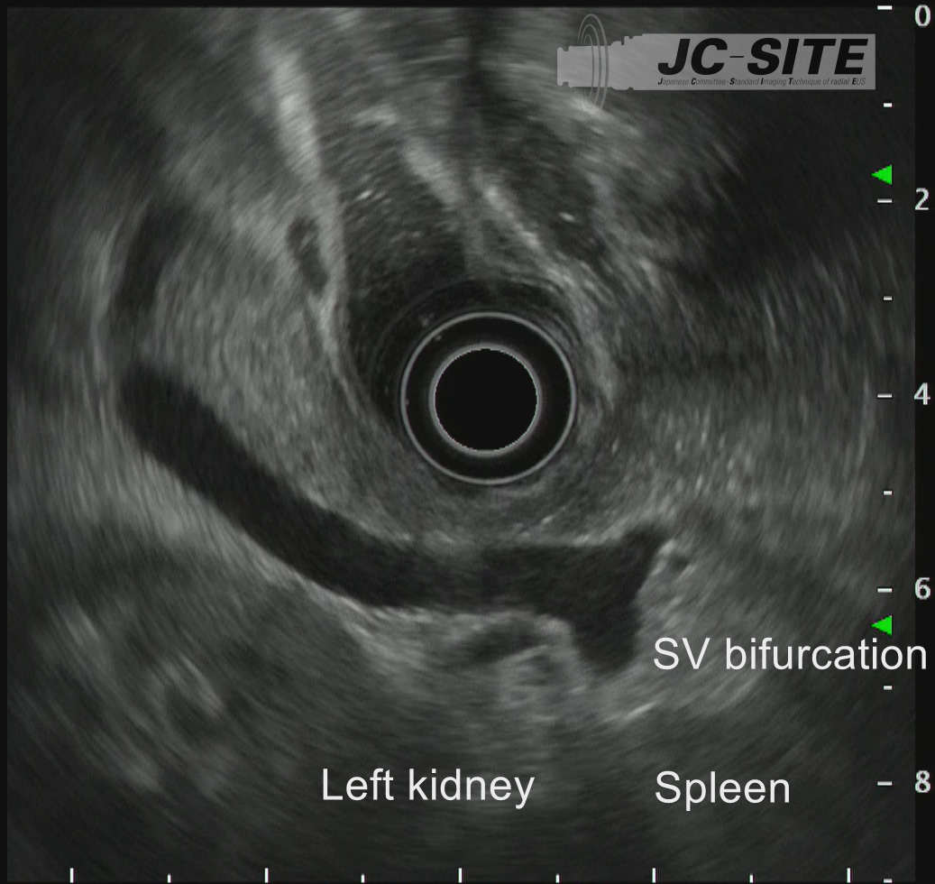
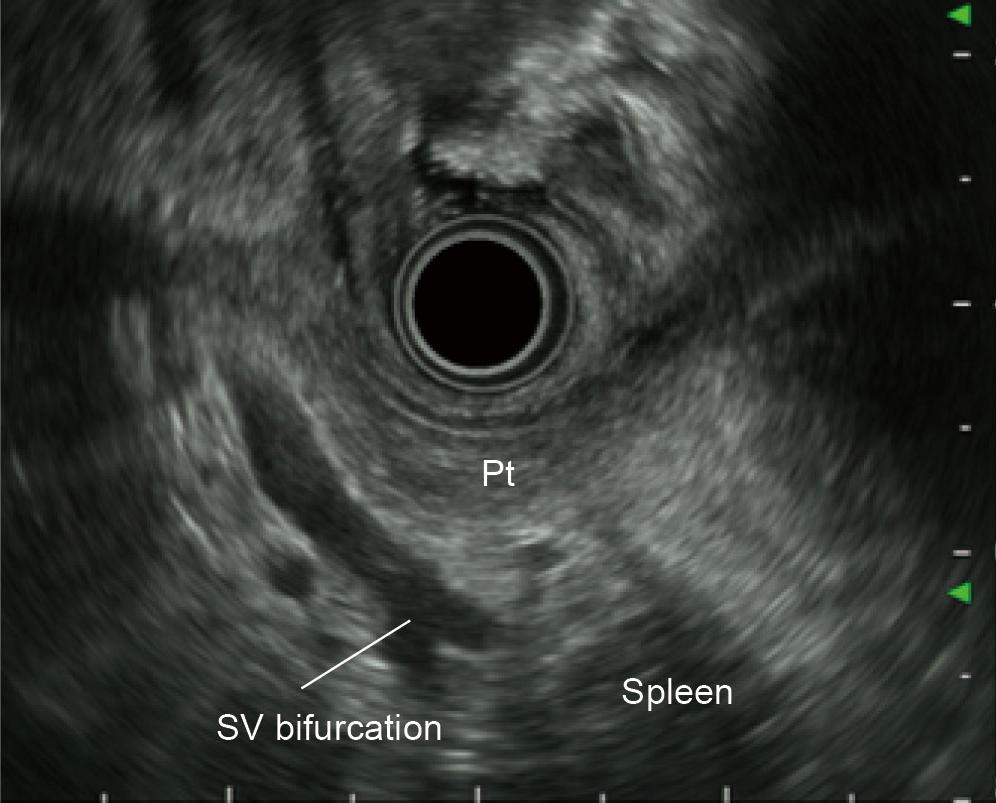
STEP 3 | Tail of the pancreas/hilum of the spleen
Rotate the scope further clockwise. Then use the splenic vein bifurcation in the hilum of the spleen as a landmark to observe the pancreatic tail.

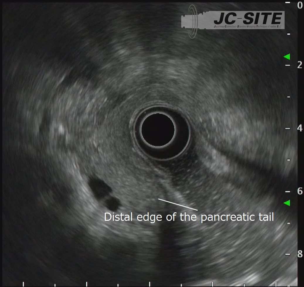
STEP 4 | Distal edge of the pancreatic tail
While following the pancreatic parenchyma, advance the scope a little to observe the distal edge of the pancreatic tail.
STEP 5 | Body of the pancreas
Rotate the scope counterclockwise to observe the body of the pancreas again.

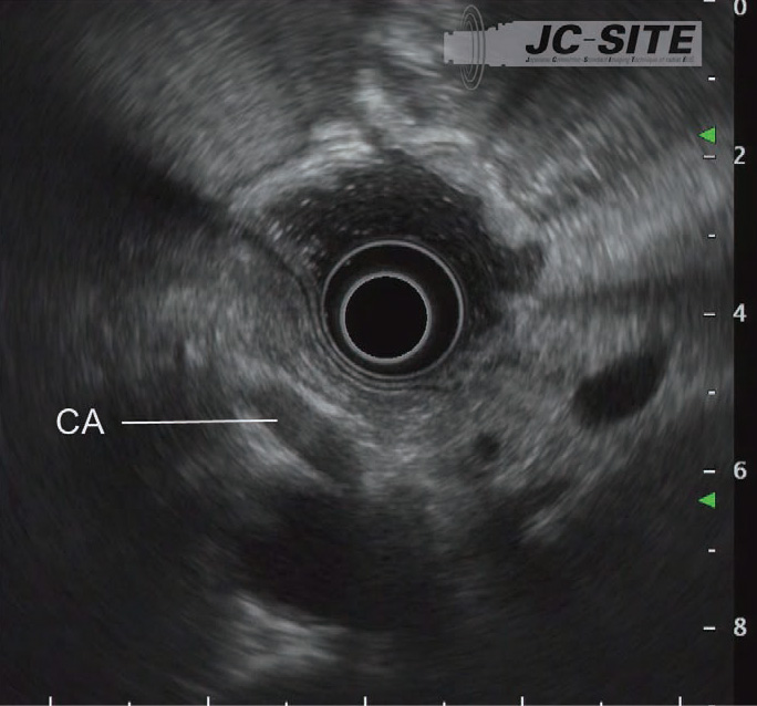
STEP 6 | Celiac artery
After observing the pancreatic body, pull the scope slightly to image the celiac artery branching off from the aorta. Check whether there is a swollen lymph node in the surroundings.

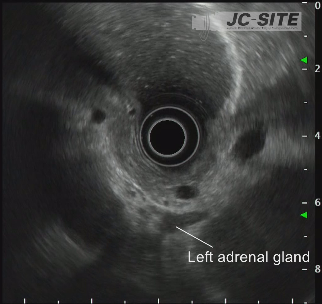
STEP 7 | Left adrenal gland
The left adrenal gland is visualized between the left kidney and the aorta.

 ③
③How to find the pancreas (the body and tail of the pancreas) via the stomach
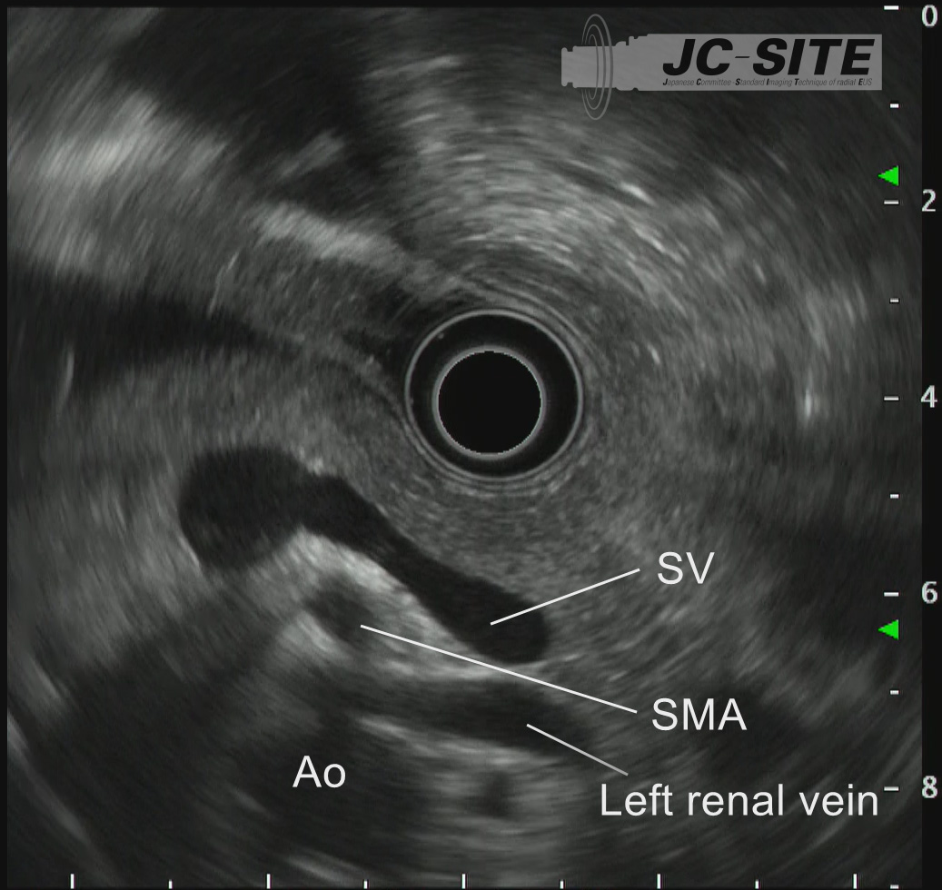
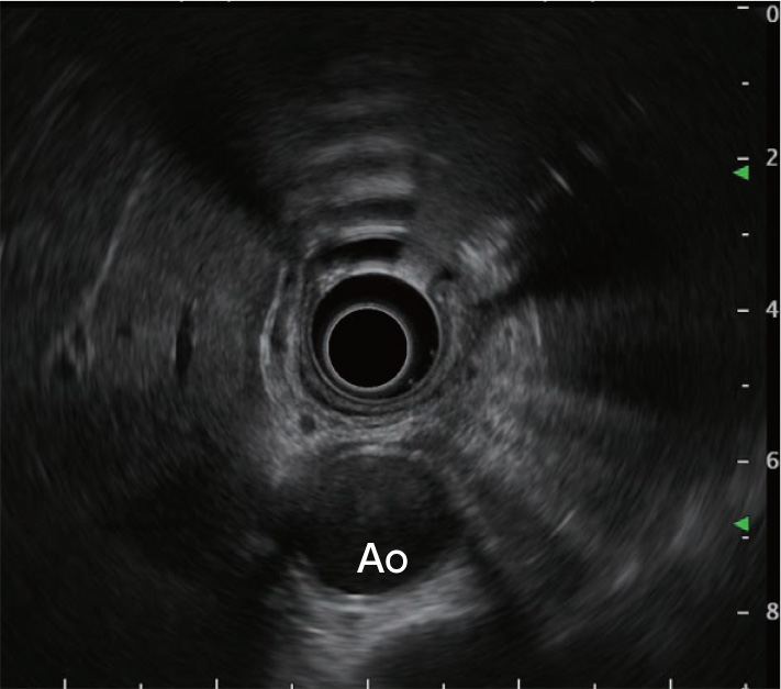
The pancreas is sometimes difficult to visualize due to pancreatic atrophy or fatty replacement (fatty infiltration). Should this be the case, pull the scope as close as possible to the esophagogastric junction in transgastric operation so that a cross-sectional image of the aorta appears right below the transducer. Rotate the scope until this is positioned in the six o’clock direction on the screen (Fig. a). While keeping this axis, advance the scope to image the splenic vein; the pancreatic parenchyma is visualized between the splenic vein and the transducer (Fig. b). Alternatively, you can also find the left kidney by scanning the gastric body first and then image the pancreatic tail between the left kidney and the transducer (Fig. c).
 ④
④Notes on visualizing the distal edge of the pancreatic tail
When visualizing the distal edge of the pancreatic tail, basically start by scanning in the direction of the pancreatic tail along the splenic artery and vein and follow these vessels until they bifurcate. Here, you should closely examine the distribution of the pancreatic parenchyma. The pancreatic parenchyma is distributed in a number of ways. These include: lying along the splenic artery and vein; heading away from the splenic artery and vein towards the distal side of the patient and entering in the space between the left kidney and the spleen (the pancreatic parenchyma appears far from the transducer when visualized, Fig. a); and leading to the splenic side away from the splenic artery and vein towards the proximal side of the patient (the pancreatic parenchyma appears close to the transducer when visualized, Fig. b).
When a beginner is going to visualize the pancreatic tail, it is a good idea to image the region with CT or MRI prior to EUS to understand the distribution of structures of the pancreatic tail region. The important thing is not to miss lesions present in the distal edge of the pancreatic tail.

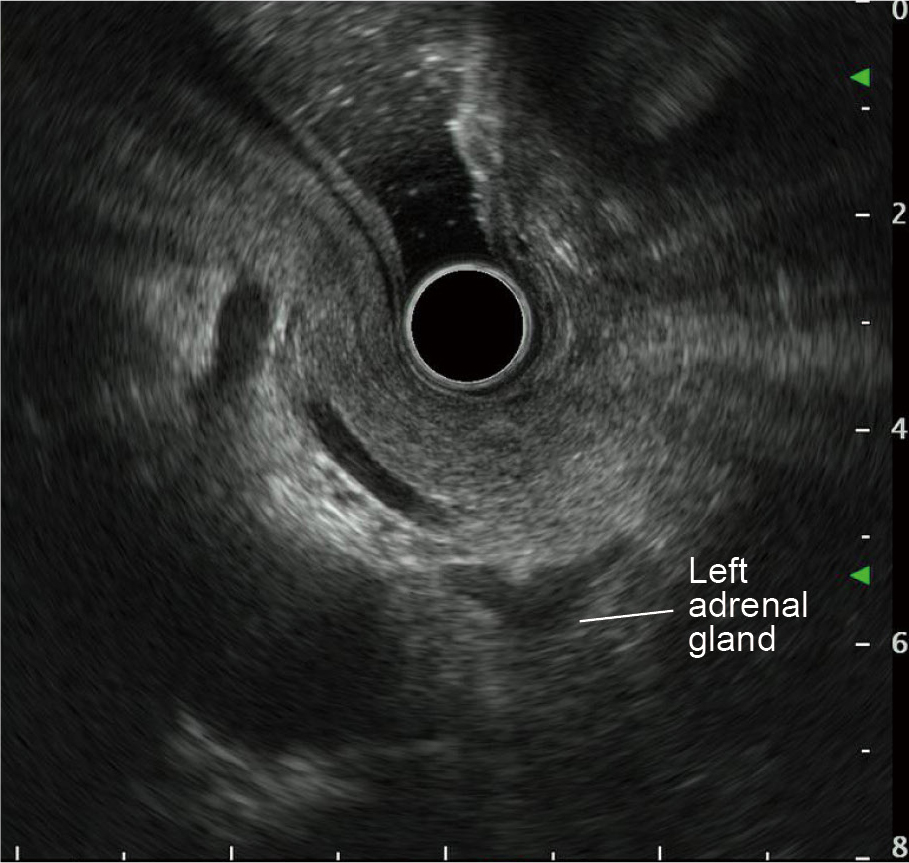
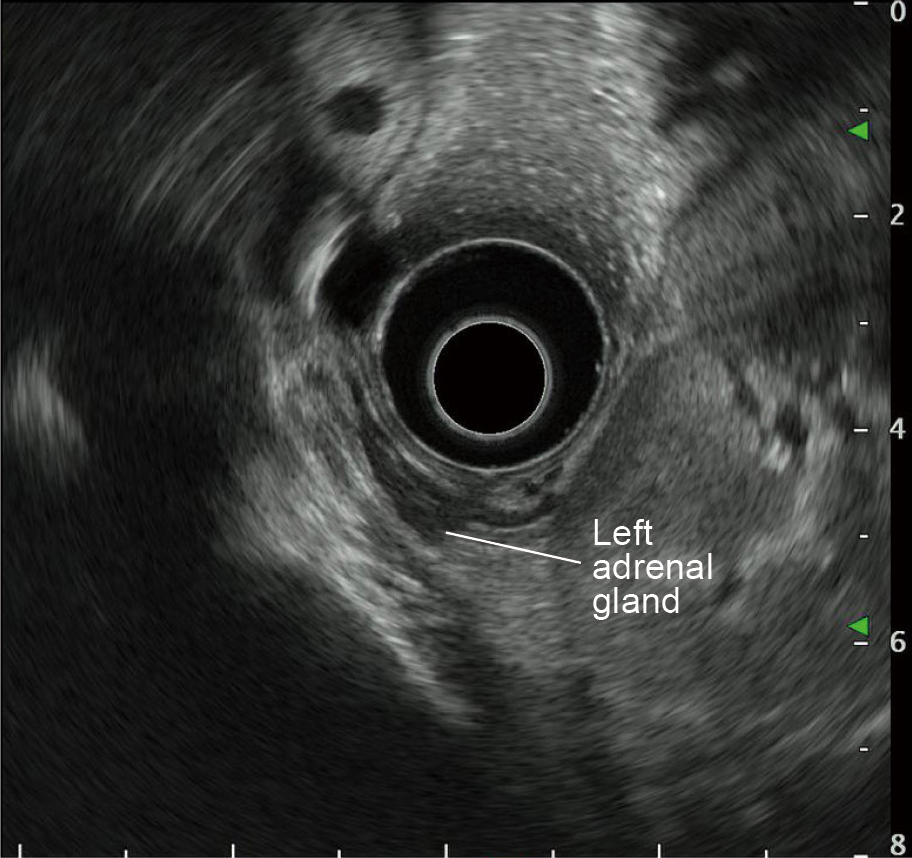
Observing the left adrenal gland How-to
The left adrenal gland can also be observed with radial EUS in the same manner as convex (linear) EUS. Displaying the “seagull sign” in the EUS image, the left adrenal gland is positioned in the upper left of the left kidney on the dorsal side of the pancreatic tail. It can either be displayed simultaneously on the dorsal side of the pancreatic tail when the pancreatic tail is observed (Fig. a) or displayed between the left kidney and the aorta when the scope is pulled while the left kidney is observed (Fig. b).
- Content Type