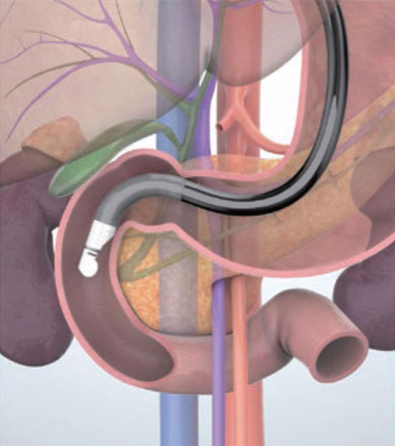
Imaging Techniques Descending part of the duodenum approach
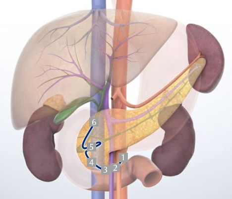
1 Aorta
2 Horizontal part of the duodenum/head of the pancreas
3 Superior mesenteric vein/neck of the pancreas
4 Inferior head of the pancreas
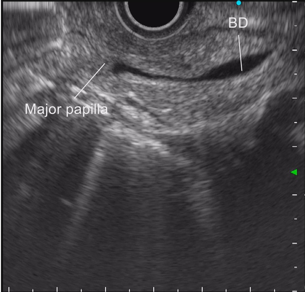
5 Major papilla; main pancreatic duct
6 Bile duct
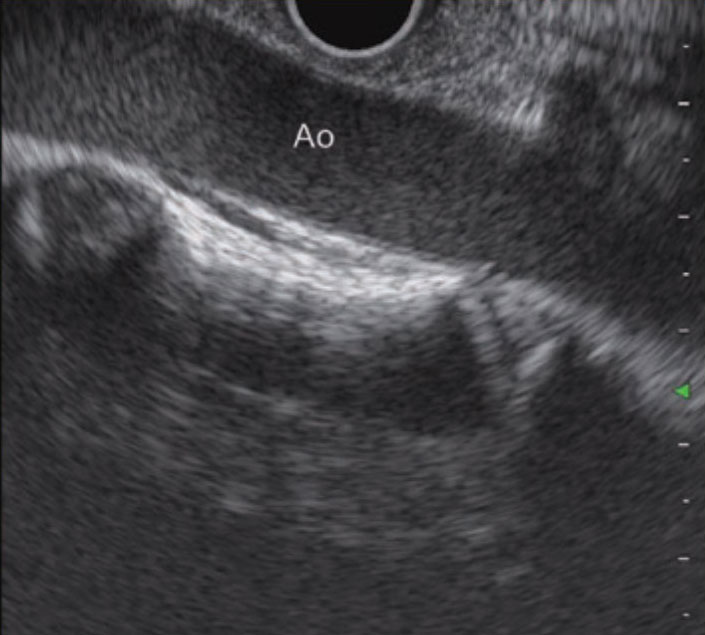
STEP 1 Aorta
In the descending part of the duodenum, start imaging in a short scope position in which the scope is stretched. When the scope is turned clockwise, the descending aorta is imaged in the long axis direction.

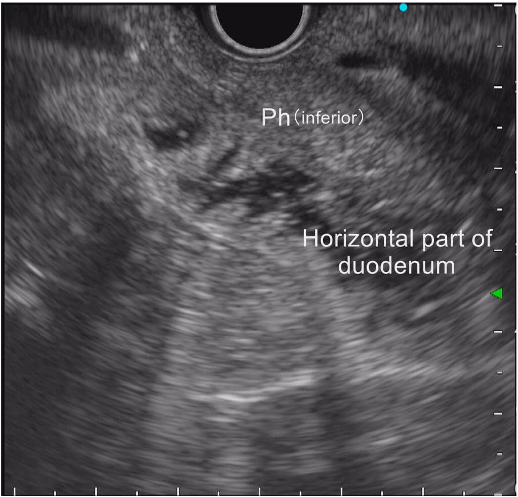
STEP 2 Horizontal part of the duodenum/head of the pancreas
Keep turning the scope clockwise to image the horizontal part of the duodenum and the inferior head of the pancreas. Inject lukewarm water to make the horizontal part of the duodenum easier to identify.
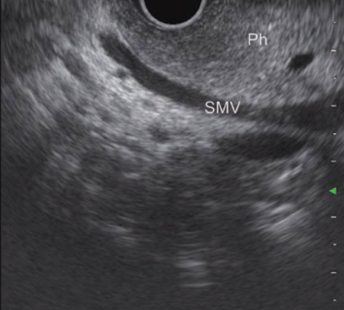
STEP 3 Superior mesenteric vein/head of the pancreas
While withdrawing the scope slightly, turn it clockwise a little at a time to image the long axis of the superior mesenteric vein. Carefully observe the head of the pancreas, which will be visible between the superior mesenteric vein and the transducer.

STEP 4 Inferior head of the pancreas
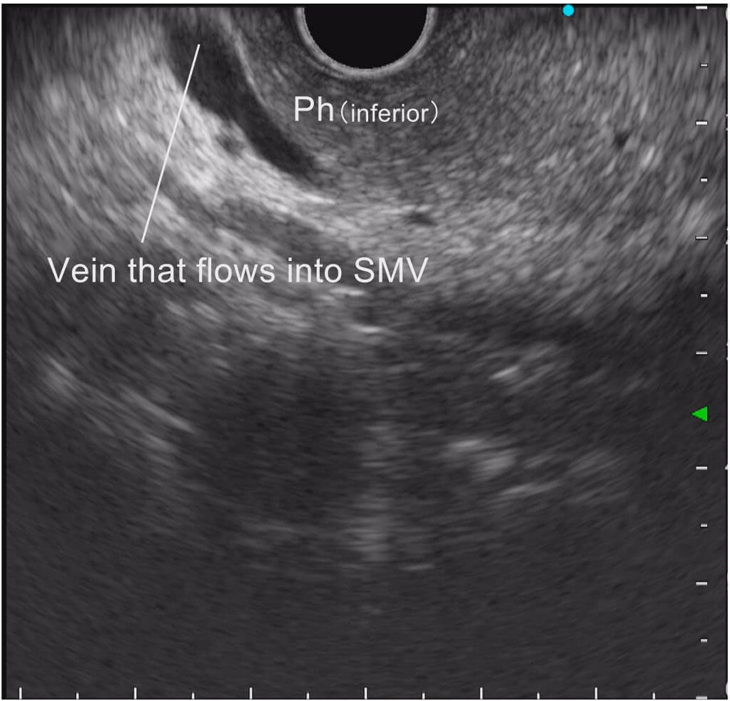
To make sure you don’t miss the inferior head of the pancreas, try to follow the superior mesenteric vein towards the inferior side. Whenever a vessel (such as the pancreaticoduodenal vein) that flows into the superior mesenteric vein is imaged, observe carefully.
STEP 5-1 Major papilla
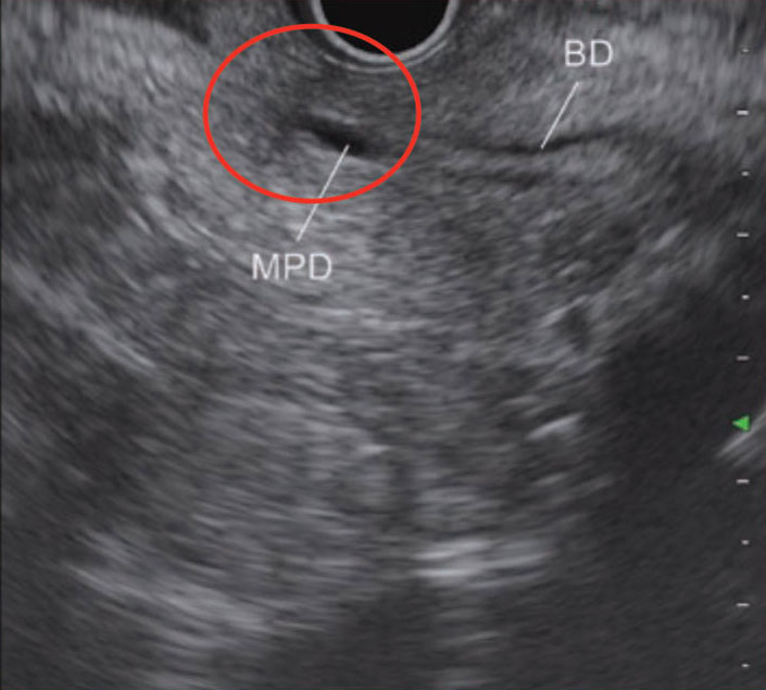
As you observe the head of the pancreas while slowly withdrawing the scope, two luminal structures will be visualized. You should be able to identify the major papilla (circled section) on the left side of the image (see “Tip 8”). The bile duct is on the proximal side and the main pancreatic duct is on the distal side. Injecting lukewarm water will expand the descending part of the duodenum, facilitating observation.

STEP 5-2 Main pancreatic duct
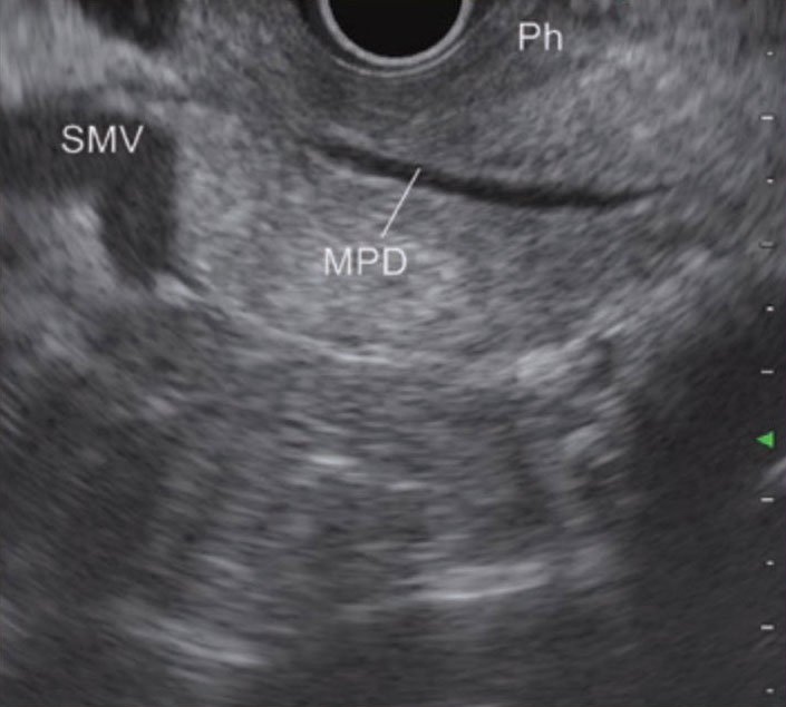
After identifying the area of the major papilla (bile duct/pancreatic duct), turn the scope a little bit clockwise. The main pancreatic duct will be imaged in the long axis direction.
STEP 6 Bile duct
Turn the scope counterclockwise while viewing the main pancreatic duct. Follow the bile duct towards the side of the hepatic hilum while withdrawing the scope to observe as far as the extrahepatic bile duct. Be careful not to pull the scope out. (see “Tip 9”).
Tips for imaging the inferior head of the pancreas and the major papilla from the descending part of the duodenum (See steps 4 & 5 in “Descending part of the duodenum approach”.)
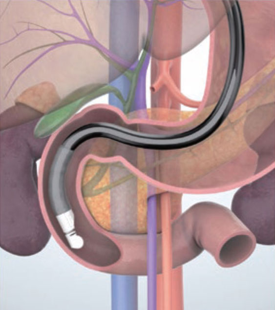
To properly observe the inferior head of the pancreas, maneuver the scope in the descending part of the duodenum so that it is in the short scope position and the transducer is positioned near the inferior duodenal angulus. From this position, observe the entire inferior head of the pancreas by withdrawing the scope while frequently using the axial rotation of the scope. Injecting deaerated water warmed to about body temperature will make it easier to see how the horizontal part of the duodenum extends and to recognize the position of the inferior head of the pancreas in the ultrasound image (Fig. a).
In this position, you can image the superior mesenteric vein and the superior mesenteric artery located on the distal side of the vein by turning the scope clockwise. This will enable you to confirm the inferior head of the pancreas visualized at somewhat high echo in between the superior mesenteric vein and the transducer. Next, turn the scope counterclockwise to observe as far as the aortic side. Then, while withdrawing the scope slightly, image the main pancreatic duct and the bile duct sequentially. Also, image the major papilla located at the distal end of these ducts.
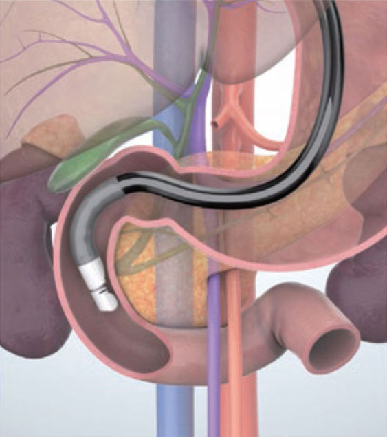
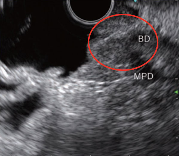
As soon as the major papilla has been imaged, it is important that you observe the anterior side of the inferior head of the pancreas by turning the scope clockwise. Also, when observing the duodenal major papilla, either suction all the air around the transducer or inflate the balloon with water to eliminate any air between the major papilla and the transducer. Injecting and filling up the descending part of the duodenum with deaerated water will give you a clear view of the structure of the major papilla (circled section), allowing you to perform detailed observation (Fig. b).

How to keep the scope from coming out of the descending part of the duodenum (See step 6 in Descending part of the duodenum approach”.)
In some cases, the scope can come out very easily during observation from the descending part of the duodenum in the short scope position, preventing you from properly observing the major papilla and the inferior head of the pancreas. To avoid this, turn the scope hard in the clockwise direction and steeply angulate the scope tip upwards. Try to keep the scope this way, moving it as little as possible during observation. Another way of keeping the scope from coming out is to inflate the balloon.
If the scope still looks like it’s going to come out, confirm the major papilla in the endoscopic image, then bring the transducer in contact with the major papilla, and start imaging from the major papilla. Doing this may allow you to properly observe the major papilla before the scope comes out. If you do this, you would observe the descending part of the pancreas after observing the major papilla.
Imaging the uncinate process of the pancreas
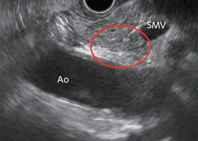
The uncinate process of the pancreas is an extension of the head of the pancreas and refers to a hook-shaped projection protruding from the inferior head of the pancreas to the left posterior side of the superior mesenteric vein. The notch created by the uncinate process of the pancreas sticking out of the head of the pancreas is called the pancreatic incisure, through which the superior mesenteric vein passes. Consequently, the area around the uncinate process of the pancreas is generally imaged between the superior mesenteric vein and the aorta.
To observe the entire head of the pancreas without missing anything, it is necessary that you understand the anatomical positioning of the uncinate process of the pancreas and carefully observe it and the surrounding area. From the descending part of the duodenum, you can recognize the area of the uncinate process of the pancreas by first visualizing the superior mesenteric vein and the head of the pancreas and then turning the scope counterclockwise from there to observe the aortic side. On the other hand, if you have observed the head of the pancreas by visualizing the aorta in the long axis direction while withdrawing the scope, you can image the area of the uncinate process of the pancreas by turning the scope clockwise from there (Fig., circled section).
- Content Type