Colorectal Case 1
Prof. Stefan Seewald
GastroZentrum Hirslanden, Zurich, Switzerland
Scope:CF-EZ1500DI
Case: Sessile serrated adenoma
Organ: Colon
Patient information: M, 60s
Medical history: Preventive colonoscopy
1. WLI Overview
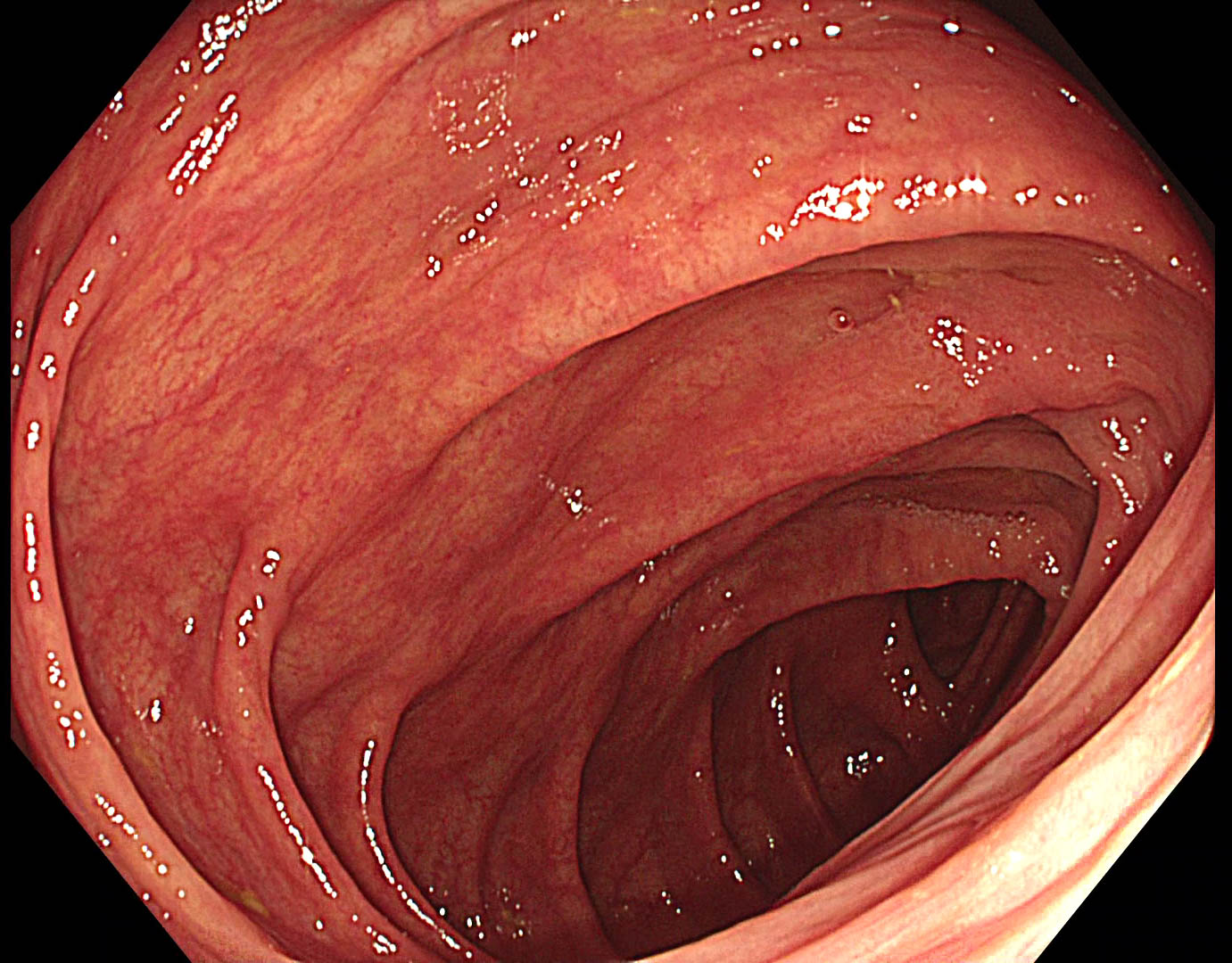
Thickening of fold indicates a lesion extending on a fold at 2 o’clock
2. TXI 1 Overview
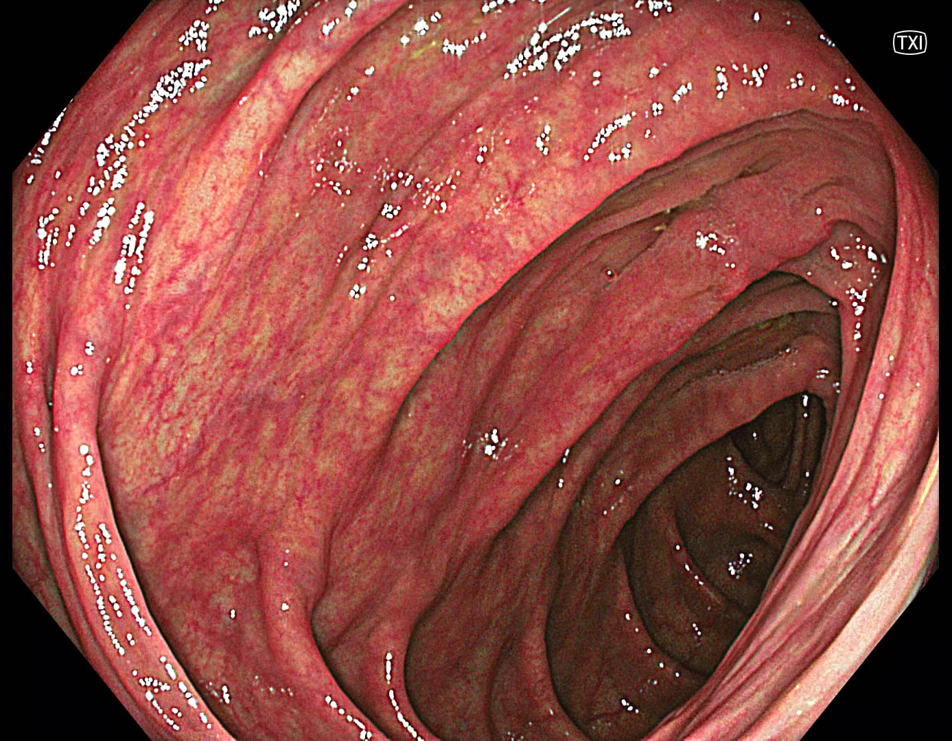
TXI increases brightness and color contrast. However, visibility of the lesion is not amplified sufficiently.
3. NBI Overview
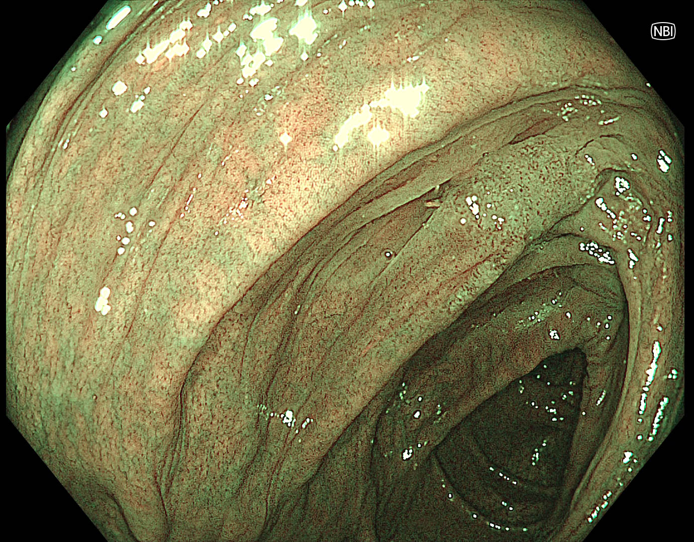
With NBI, the color contrast between healthy background mucosa and the lesion is most comprehensive in this case.
4. Approach with NBI
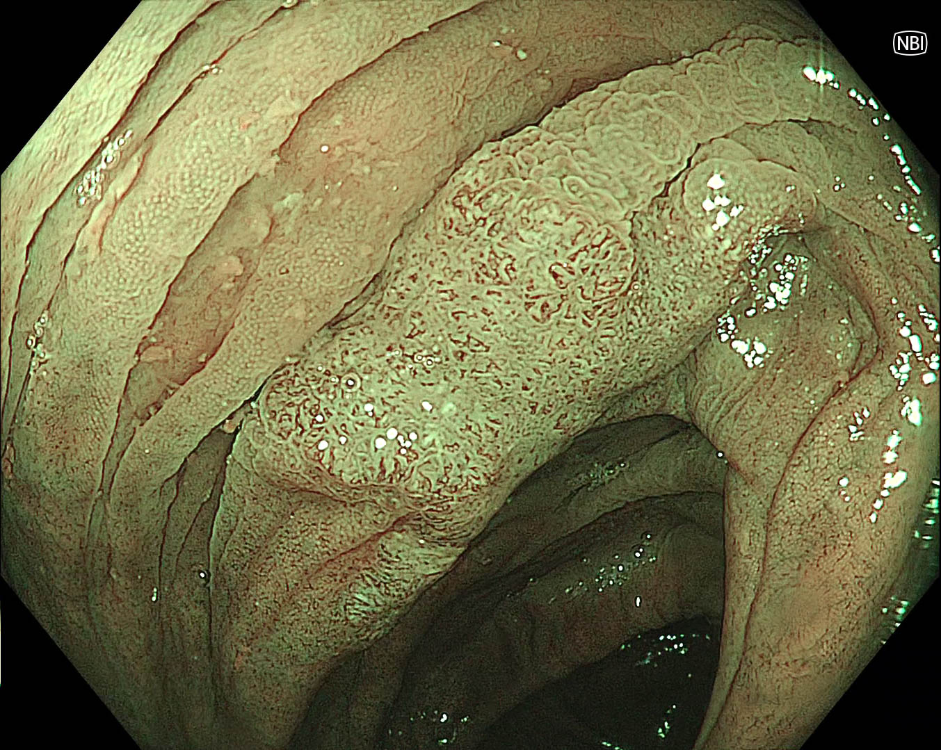
With NBI, we can observe areas with high and low vessels density and irregular pattern.
5. Near Focus mode with NBI
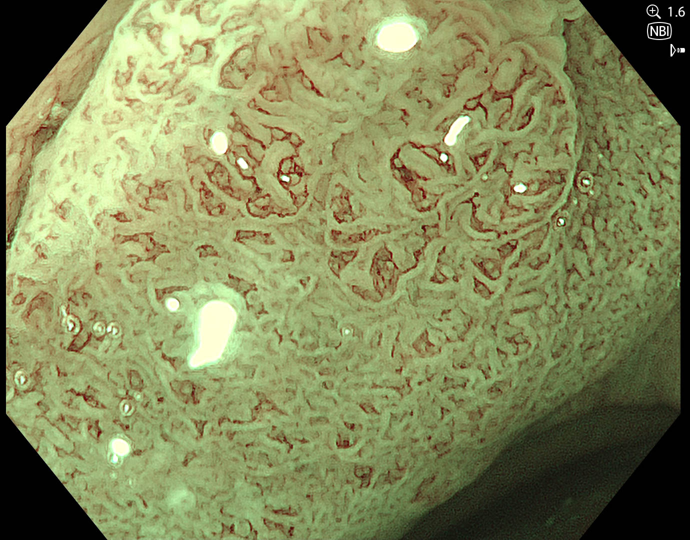
Near Focus mode + 1.6x electronic zoom reveals adenoma with high degree of irregularity (JNET 2B).
6. Near Focus mode with NBI
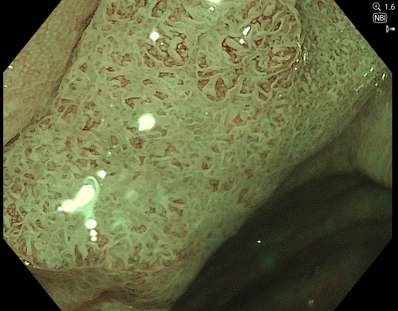
Near Focus mode + 1.6x electronic zoom reveals adenoma with high degree of irregularity (JNET 2B).
Case video
Overall Comment
In this case, NBI was most helpful to identify and characterize the lesion underlining existing evidence that it is working well if the colon is clean. EDOF and Near Focus are easy to use to characterize and predict endoscopic resectability of the lesion.
* Specifications, design and accessories are subject to change without any notice or obligation on the part of the manufacturer.
- Keyword
- Content Type