Colorectal Case 22

Prof. Yasushi Sano
Kansai Medical University, Osaka, Japan
Sano Hospital, Kobe, JapanScope: CF-EZ1500DI
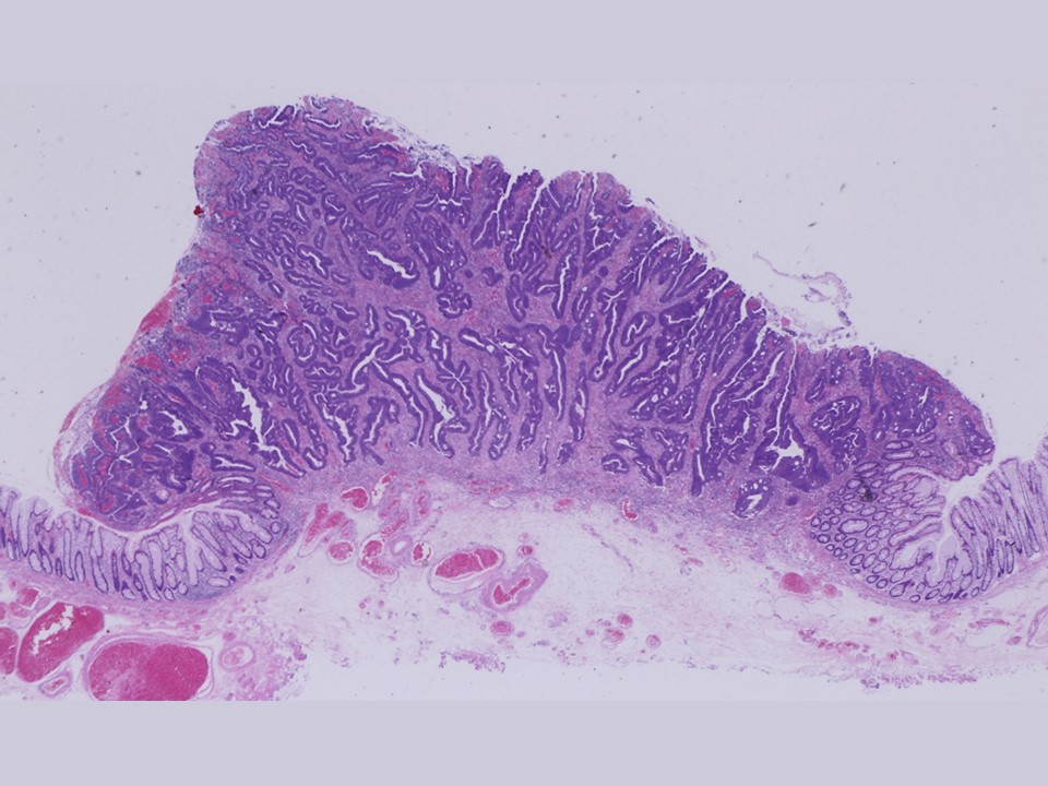
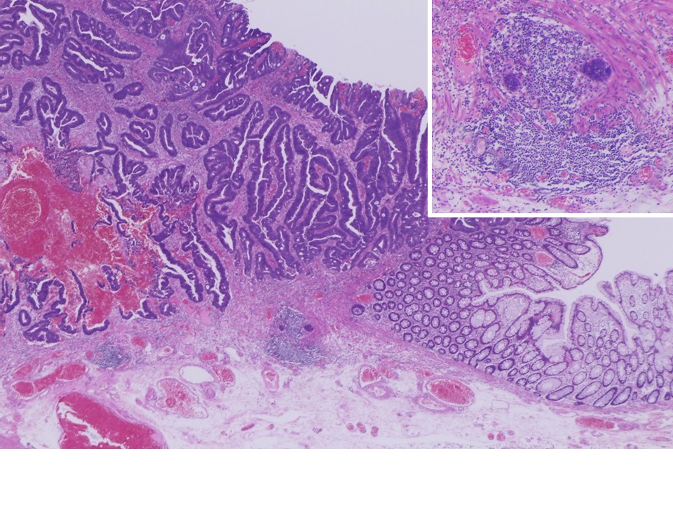
Histology: Well to moderately differentiated tubular adenocarcinoma, pT1b (SM2, 3600 µm invasion), ly0, v0, pHM0, pVM0. After ESD, no recurrence was found at 3-year follow-up.
Organ: Rectum
Patient information: F, 80s
Medical history: Melena
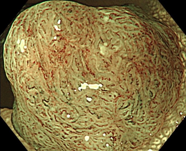
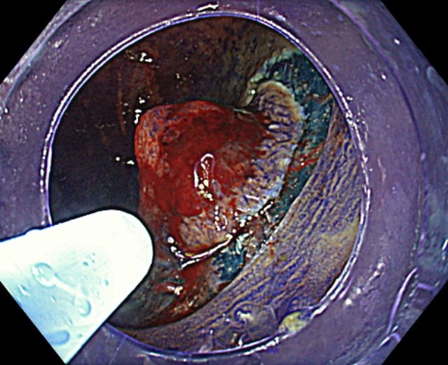
1. WL
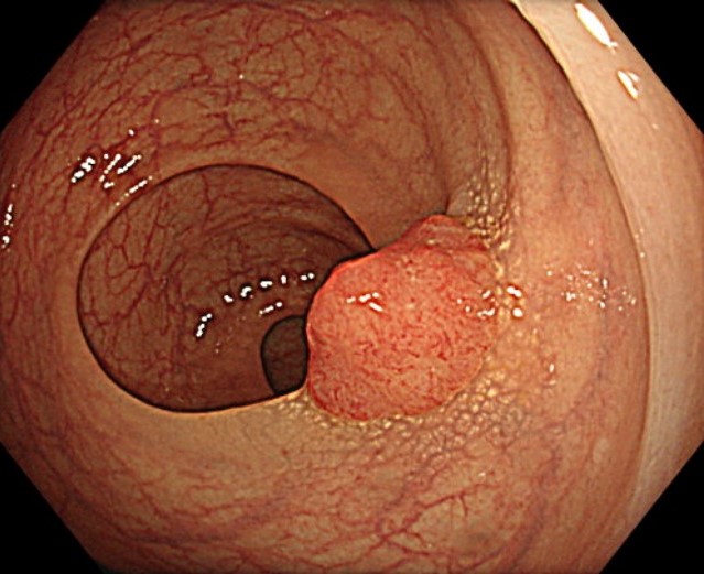
Enhancement : A8
NBI Mode : NA
TXI Mode : NA
RDI Mode : NA
BAI-MAC : On
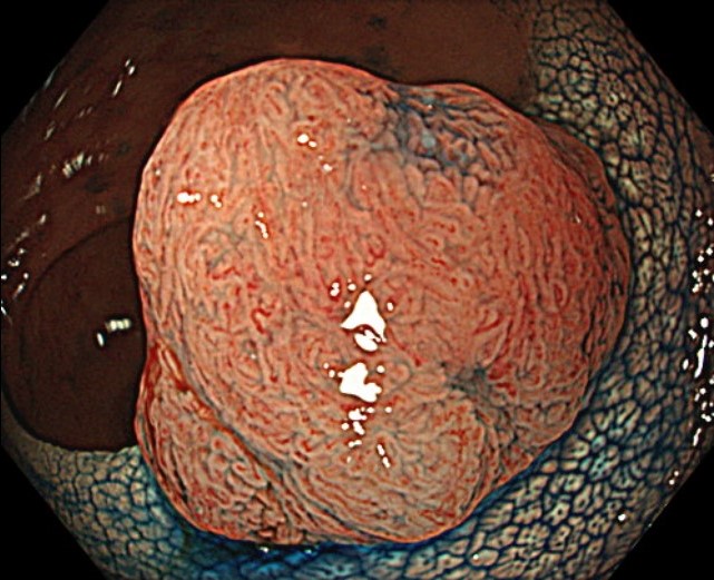
2. WL
Enhancement: A8
NBI mode: NA
TXI Mode: NA
RDI Mode: NA
BAI-MAC: On
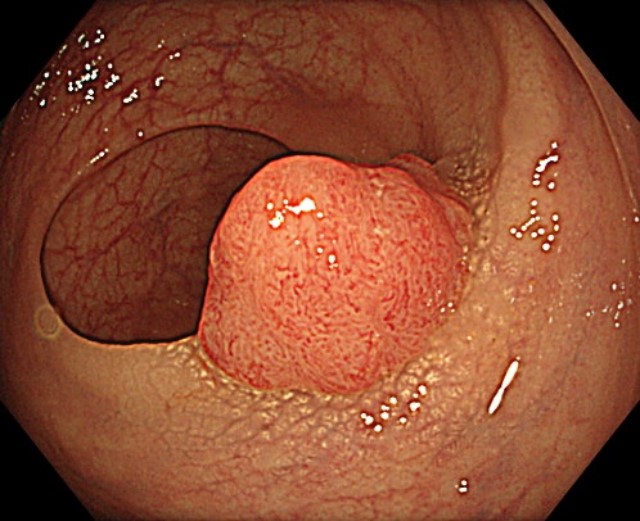
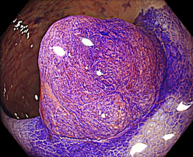
3. NBI
Enhancement : A8
NBI Mode : On
TXI Mode : NA
RDI Mode : NA
BAI-MAC : On
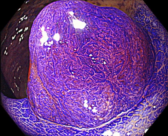
4. NBI
Enhancement : A8
NBI Mode : On (mode 3)
TXI Mode : NA
RDI Mode : NA
BAI-MAC : On
5. NBI with magnification
Enhancement : A8
NBI Mode : On
TXI Mode : NA
RDI Mode : NA
BAI-MAC : On
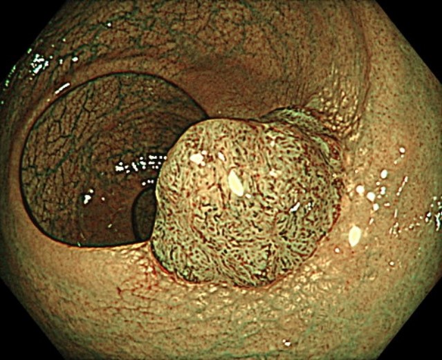
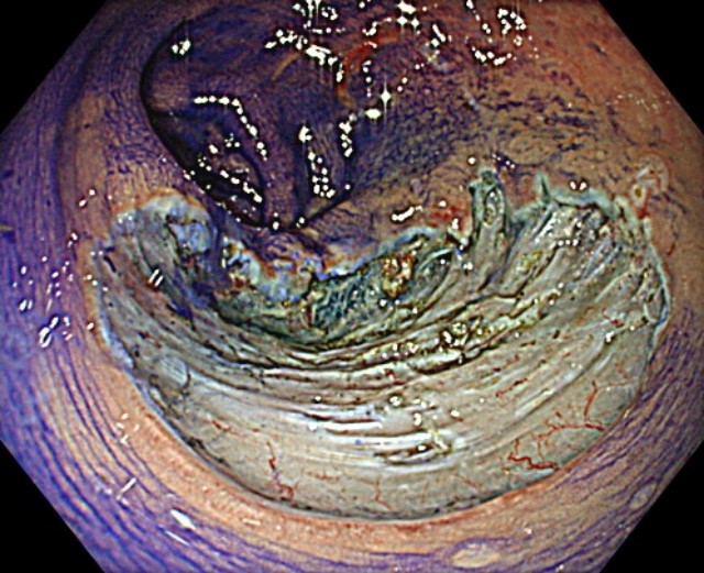
6. Chromoendoscopy
Enhancement : A8
NBI Mode : NA
TXI Mode : NA
RDI Mode : NA
BAI-MAC : On
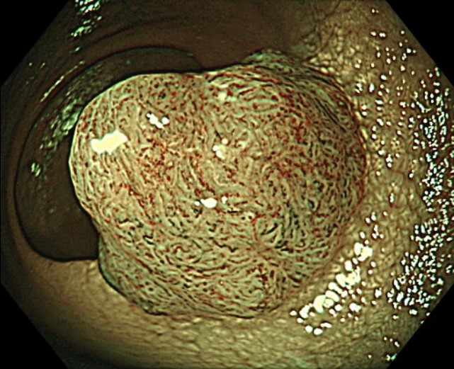
7. Crystal violet
Enhancement : A8
NBI Mode : NA
TXI Mode : NA
RDI Mode : NA
BAI-MAC : On
8. Crystal violet
Enhancement : A8
NBI Mode : NA
TXI Mode : NA
RDI Mode : NA
BAI-MAC : On
9. Endocytoscopy

Enhancement : A8
NBI Mode : NA
TXI Mode : NA
RDI Mode : NA
BAI-MAC : NA
10. ESD procedure
Enhancement : A8
NBI Mode : NA
TXI Mode : NA
RDI Mode : NA
BAI-MAC : NA
11. En bloc resection
Enhancement : A8
NBI Mode : NA
TXI Mode : NA
RDI Mode : NA
BAI-MAC : NA
12. Histology (low power)
13. Histology (high power)
Case Video
Video 1: Observation by WL, NBI
Video 2: Observation by chromoendoscopy with Indigo carmine and Crystal violet staining
Video 3: Endocytoscopy (x520)
Overall Comment
This case was an early-stage cancer (SM cancer) of type 0-Is. At first glance, it appeared to be an intramucosal carcinoma on WL, but it presented JNET 2B on NBI. In Japan, dye endoscopic observation is recommended for JNET 2B lesions 1). In this case, crystal violet observation was added, and a diagnosis of SM carcinoma was made with Vi type pits in all areas 2). Endocytoscopy also showed EC3b, which was suspicious of SM invasion 3). ESD was performed at the patient’s desire and three years have passed without evidence of recurrence.
1. Iwatate M, Sano Y, et al. Validation study for development of the Japan NBI Expert Team classification of colorectal lesions. Dig Endosc. 2018 Sep;30(5):642-651.
2. Matsuda T, Fujii T, et al. Efficacy of the invasive/non-invasive pattern by magnifying chromoendoscopy to estimate the depth of invasion of early colorectal neoplasms. Am J Gastroenterol. 2008 Nov;103(11):2700-6.
3. Kudo SE, Wakamura et al. Diagnosis of colorectal lesions with a novel endocytoscopic classification – a pilot study. Endoscopy. 2011 Oct;43(10):869-75.
* Specifications, design and accessories are subject to change without any notice or obligation on the part of the manufacturer
Prof. Yasushi Sano Case 23: Large non-polypoid rectal tumor
Prof. Han-Mo Chiu
- Keyword
- Content Type


