OLYMPUS On-Demand Library
Add to Watch
Table of Contents

Colorectal Case 23

Prof. Han-Mo Chiu

Clinical Professor, Department of Internal Medicine, College of Medicine,

National Taiwan University College of Medicine, Taiwan


Procedure Information


Scope: Diagnosis: CF-HQ290ZI; ESD: PCF-H290TI

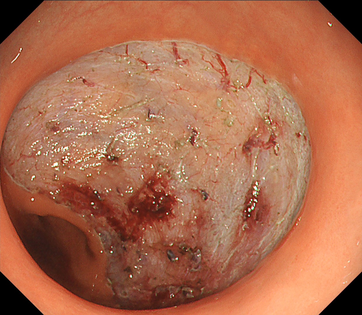
Case Findings: Large non-polypoid rectal tumor

Organ: Colorectum

Patient information: 58 years old, Male

Rectal tumor found at colonoscopy after a positive FIT (fecal immunochemical test)

Medical history: No systemic disease

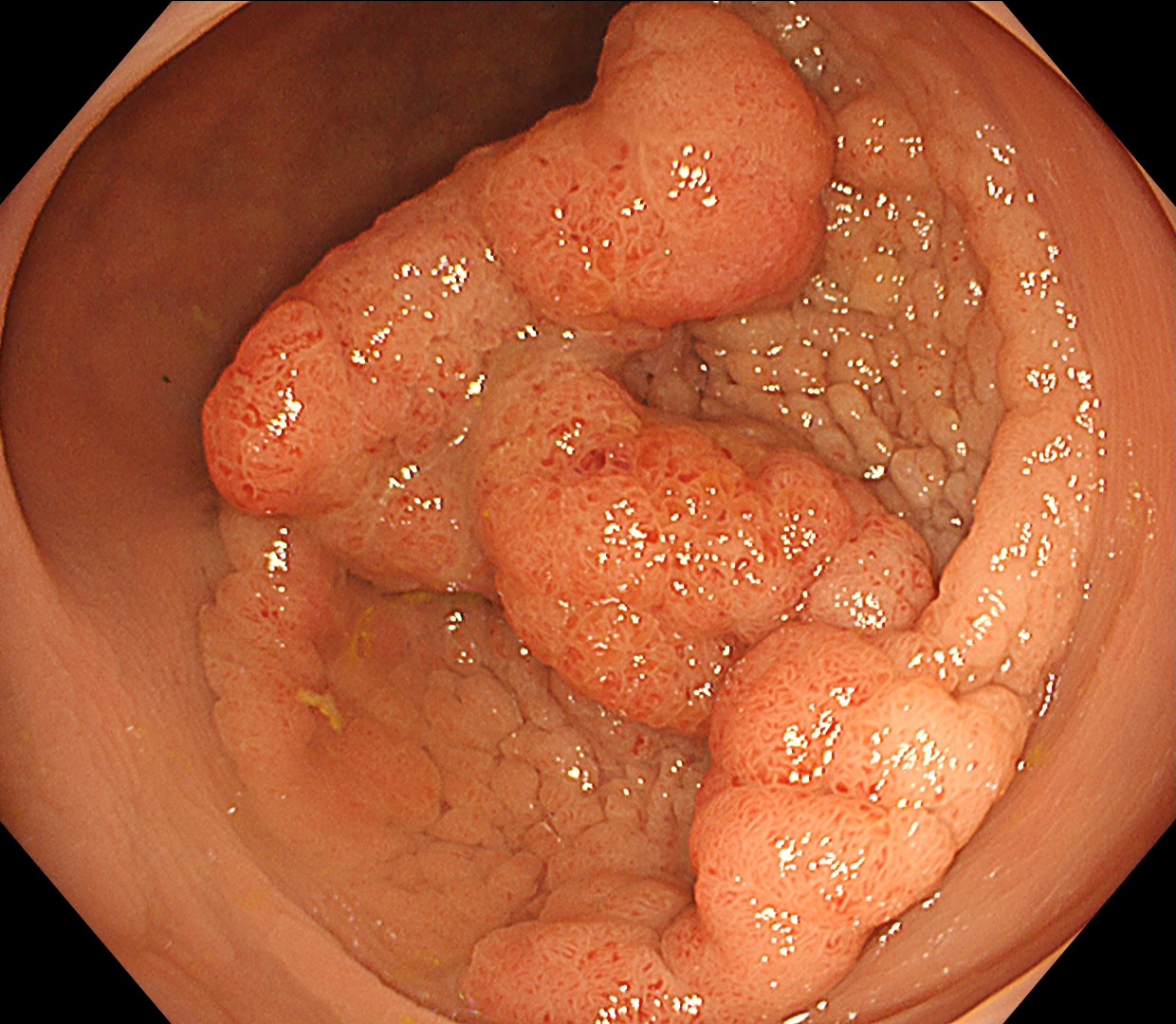
1. WLI Observation

WLE revealed a large non-polypoid tumor extending over half of the rectal circumference. The color of the large nodular parts was somewhat redder than the remining flat part.

Enhancement : A7
NBI Mode : NA
TXI Mode : NA
RDI Mode : NA
BAI-MAC : On

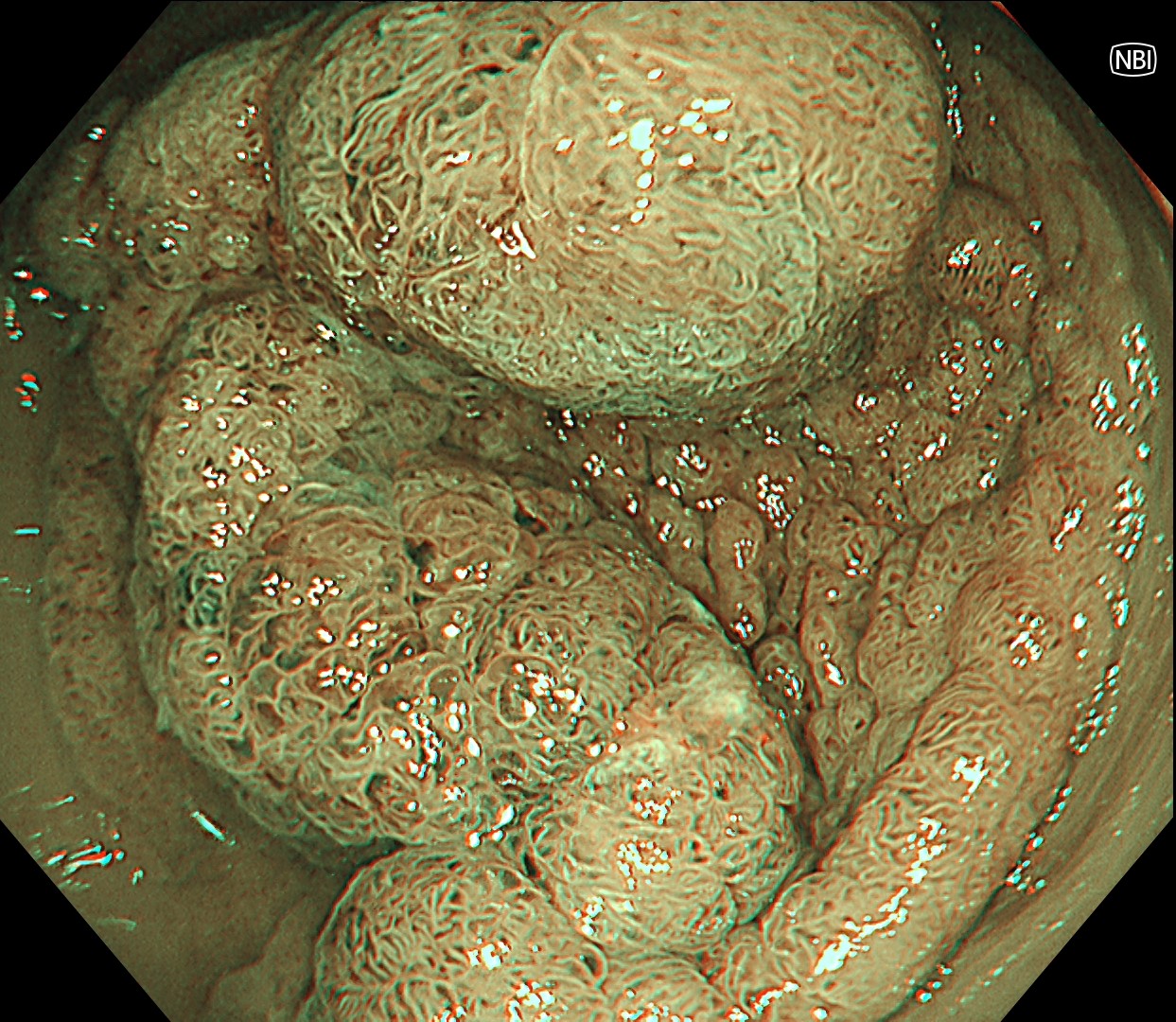
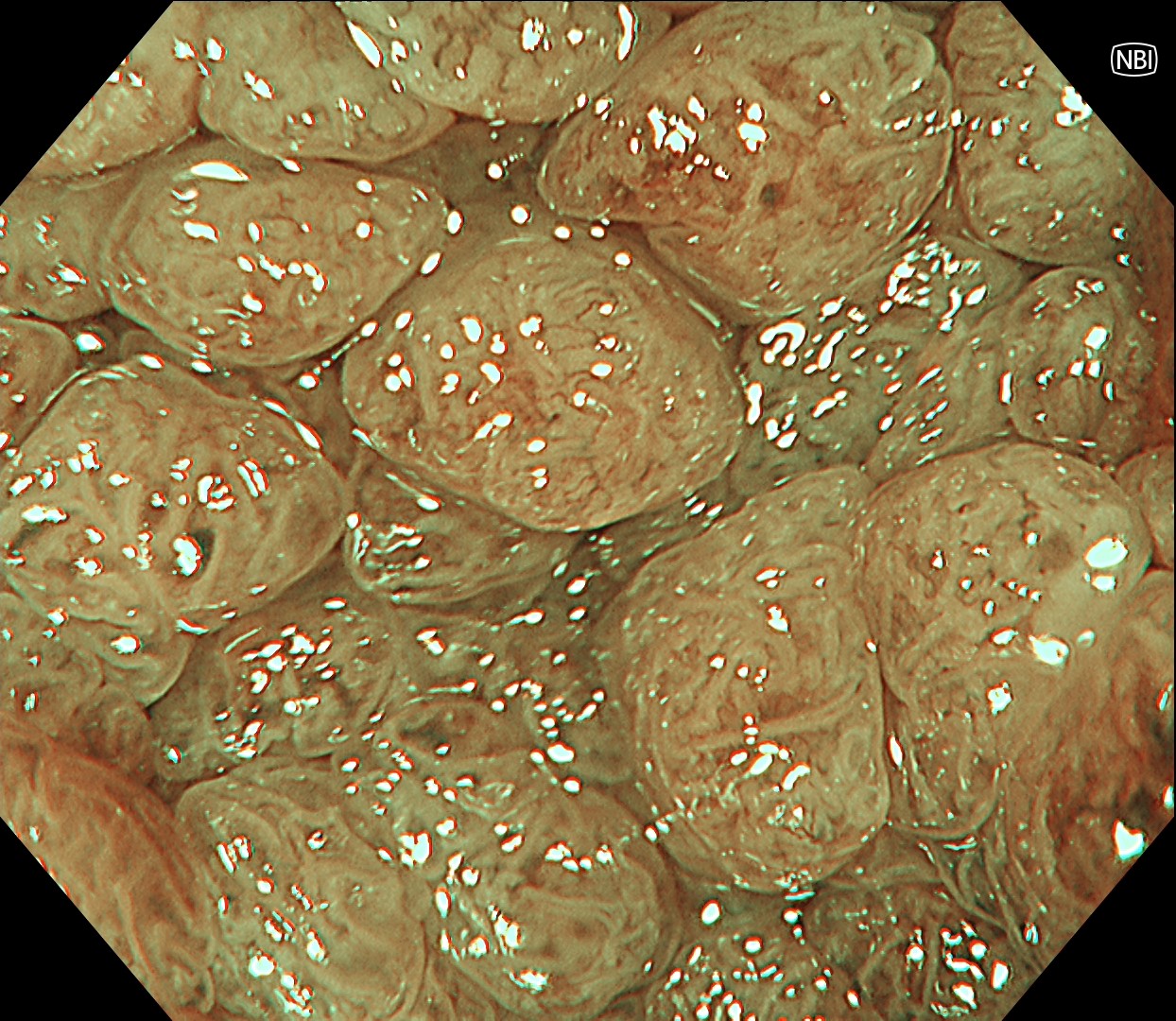
2. NBI Observation

Under NBI, the capillary network density was higher on the large nodules, with a slightly greenish area observed at the border of the nodule located at 12 o'clock.

Enhancement: A7
NBI mode: 2
TXI Mode: NA
RDI Mode: NA
BAI-MAC: On

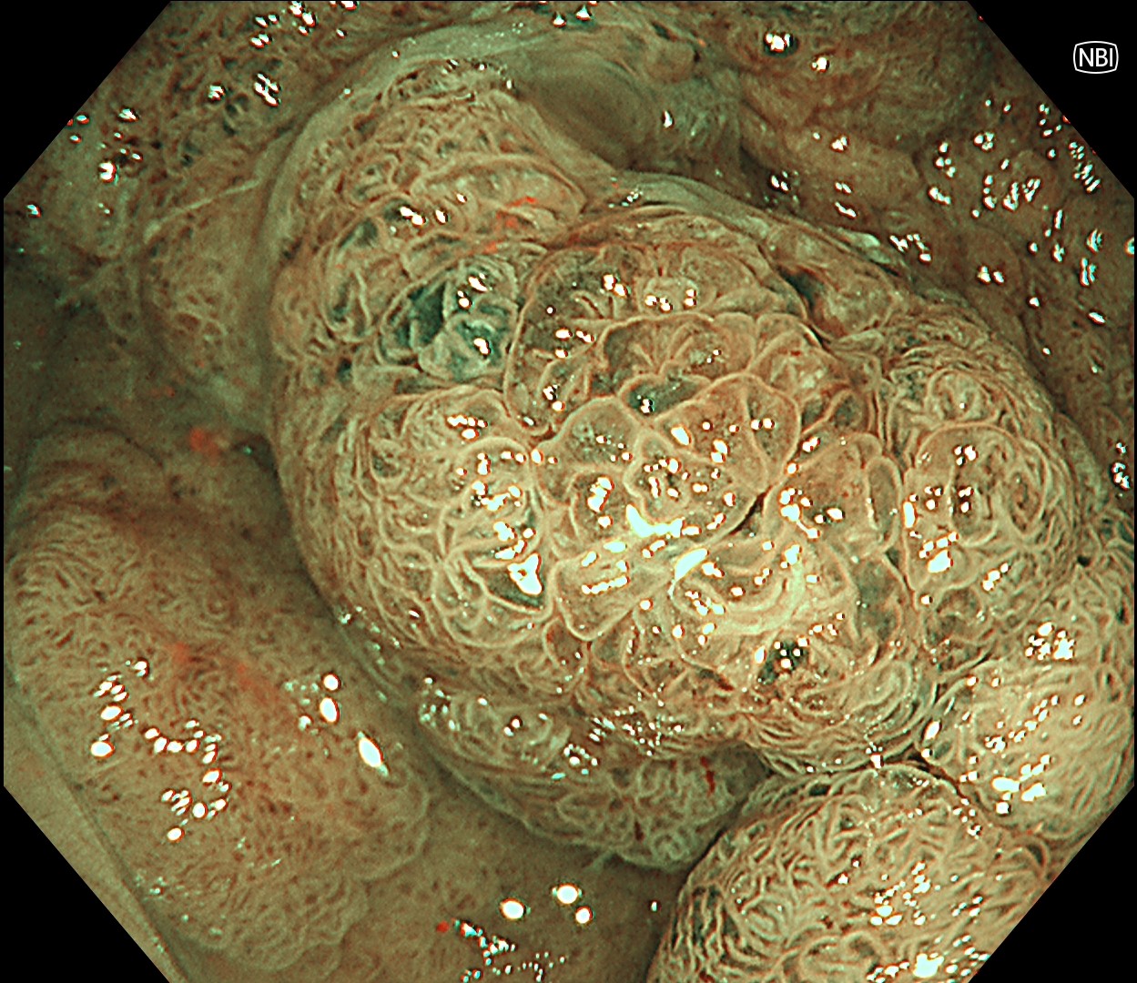
3. NBI Observation

Both surface and capillary pattern of the majority of the large nodular parts revealed regular appearance, indicating a non-invasive histology.

Enhancement : A7
NBI Mode : 2
TXI Mode : NA
RDI Mode : NA
BAI-MAC : On

4. NBI Observation

JNET 2B capillary pattern was observed focally at the margin of a large nodule (3 o’clock direction) under NBI magnification.

Enhancement : A7
NBI Mode : On
TXI Mode : NA
RDI Mode : NA
BAI-MAC : On

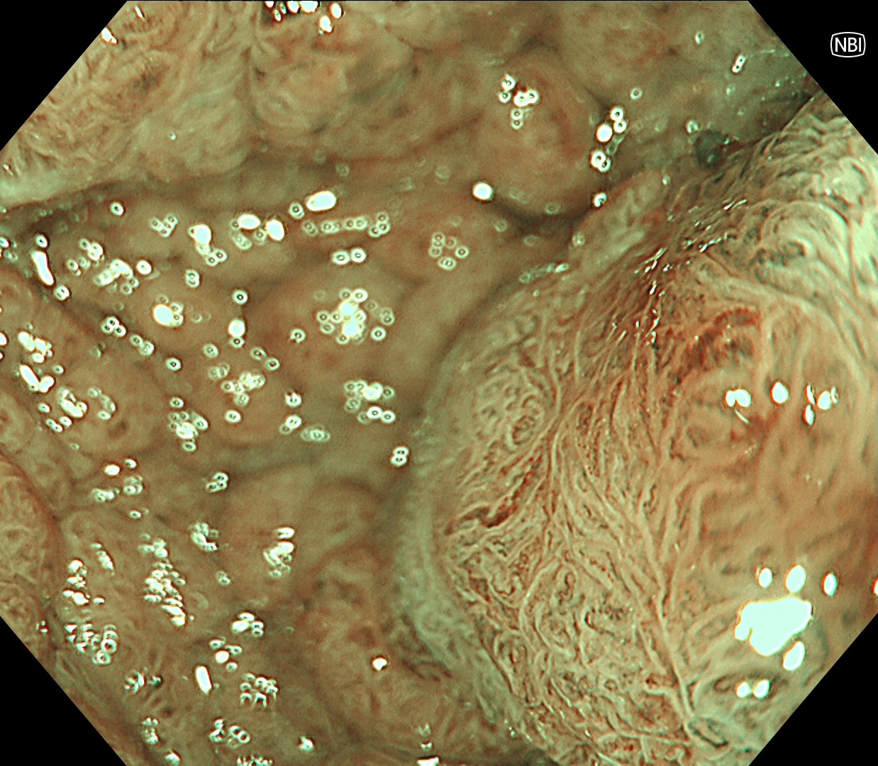
5. NBI Observation

Both surface and capillary pattern revealed regular appearance at the relatively flat and depressed area, indicating a non-invasive histology.

Enhancement : A7
NBI Mode : On
TXI Mode : NA
RDI Mode : NA
BAI-MAC : On

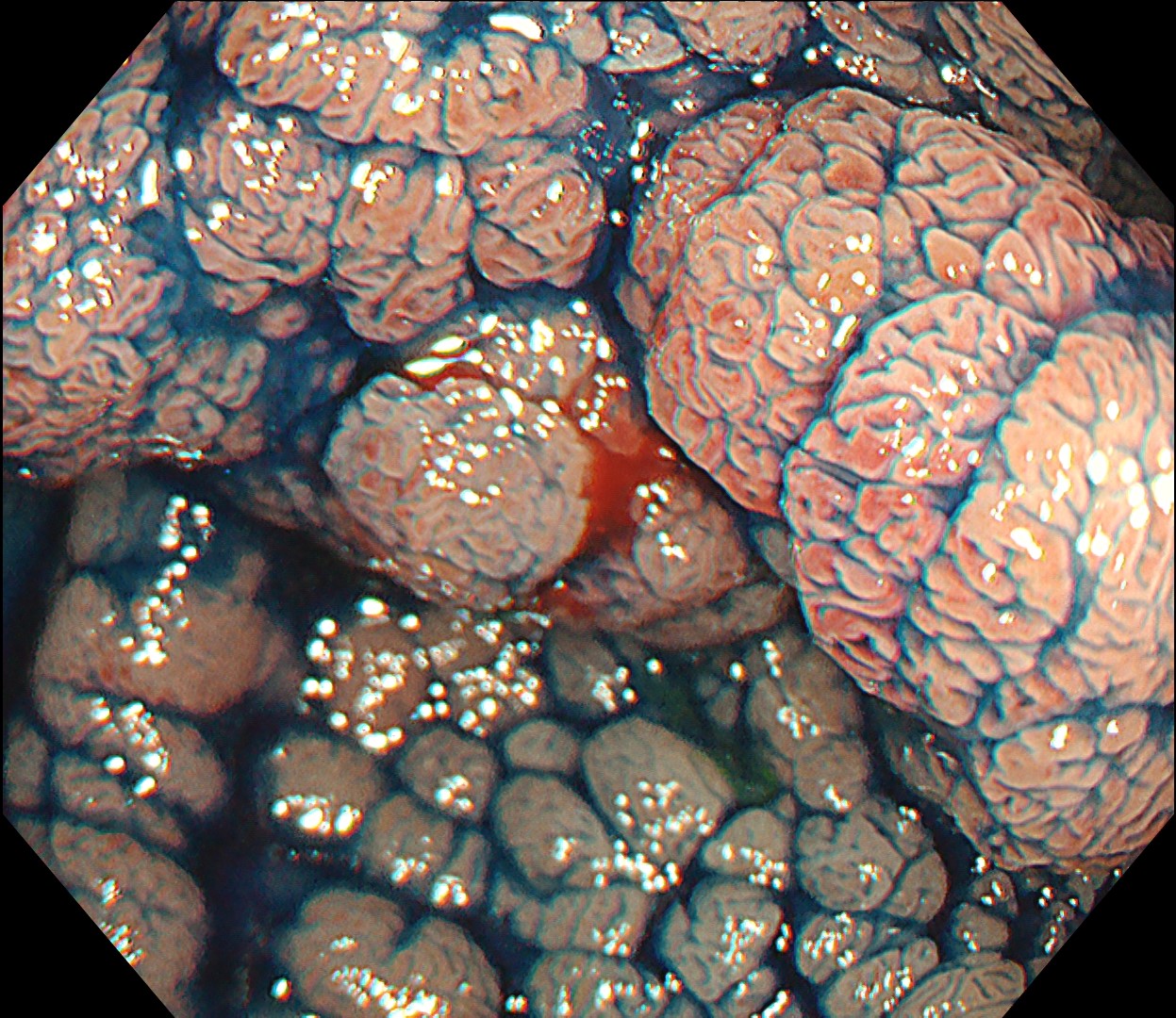
6. Chromoendoscopy

Chromoendoscopy with indigo-carmine dye spraying may help delineate the margin. It is the standard method for defining the macroscopic morphology of colorectal lesions. This lesion is morphologically interpreted as 0-Is+IIa (LST-NG-NM).

Enhancement : A7
NBI Mode : NA
TXI Mode : NA
RDI Mode : NA
BAI-MAC : On

7. Chromoendoscopy

Under macroscopic view of chromoendoscopy, the surface was mainly featured by Kudo type IV pit pattern, which indicates a non-invasive histology.

Enhancement : A7
NBI Mode : NA
TXI Mode : NA
RDI Mode : NA
BAI-MAC : On

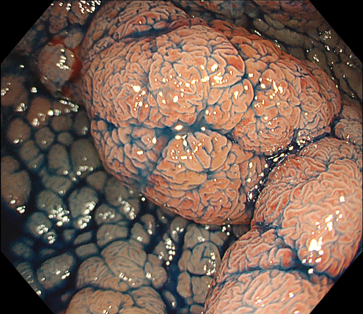
8. Chromoendoscopy

Even the surface of the large nodular portions revealed no invasive pit pattern.

Enhancement : A7
NBI Mode : NA
TXI Mode : NA
RDI Mode : NA
BAI-MAC : On

9. Chromoendoscopy
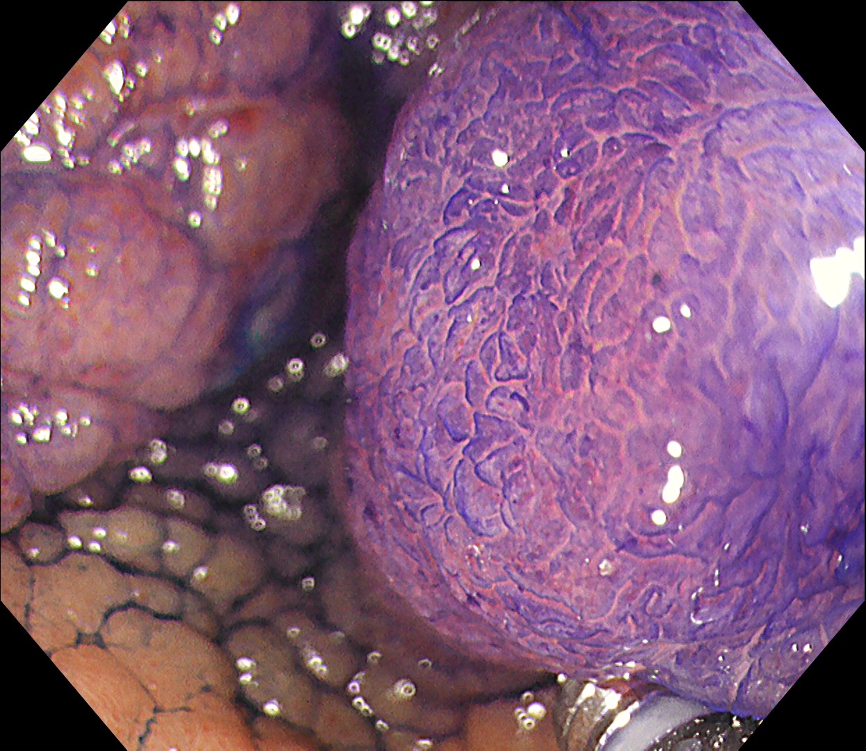
As focal JNET type 2B capillary pattern was observed at NBI magnification, chromoendoscopy with crystal violet staining was applied. The surface is mainly featured by non-invasive pit pattern, including a small focus with type Vi pit pattern at the corresponding area where JNET 2B capillary pattern was observed.

Enhancement : A7
NBI Mode : NA
TXI Mode : NA
RDI Mode : NA
BAI-MAC : On

10. ESD - incision and trimming

Under the impression of non-invasive neoplastic lesion, ESD was performed with Olympus DualKinfe J (KD-655U).

Enhancement : A7
NBI Mode : NA
TXI Mode : NA
RDI Mode : NA
BAI-MAC : On

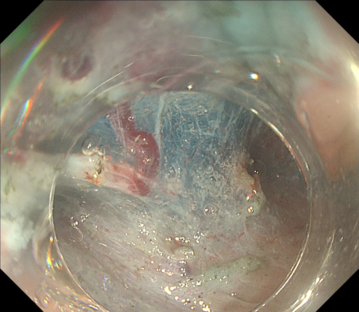
11. Submucosal dissection - 1

En bloc resection was completed.

Enhancement : A8
NBI Mode : NA
TXI Mode : NA
RDI Mode : NA
BAI-MAC : NA

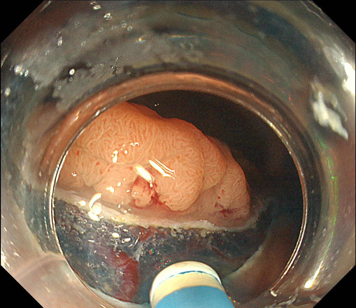
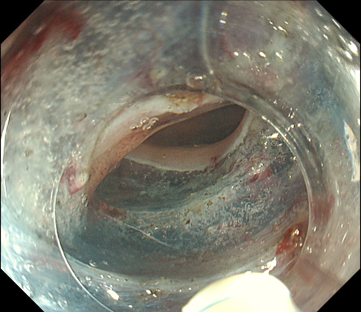
12. Submucosal dissection - 2

Tunnel was created with visible oral side of tumor

Enhancement : A7
NBI Mode : NA
TXI Mode : NA
RDI Mode : NA
BAI-MAC : On

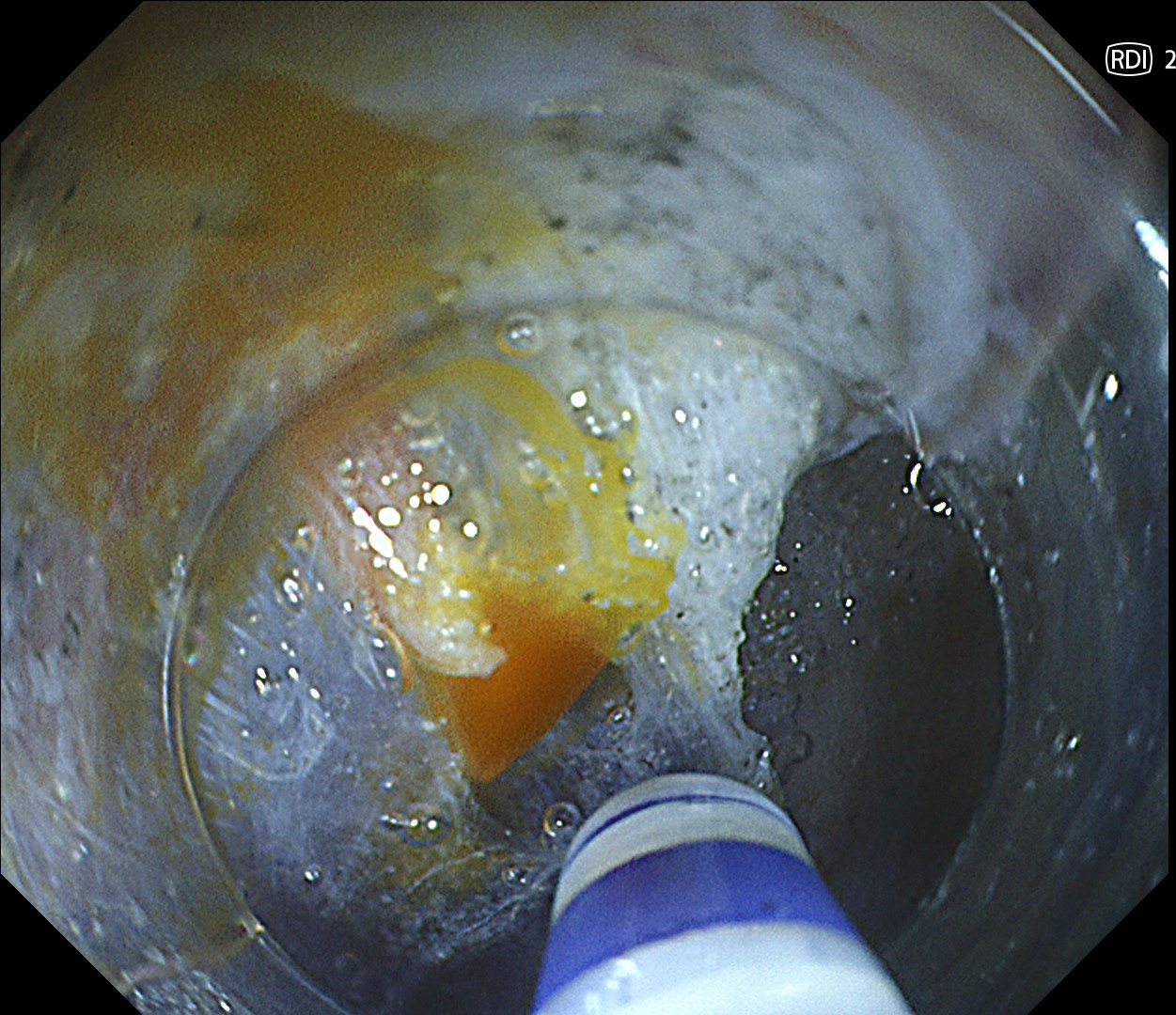
13. Hemostasis under RDI mode

Under RDI mode, identifying the bleeding vessel becomes easier, thus facilitating hemostasis.

Enhancement : A7
NBI Mode : NA
TXI Mode : NA
RDI Mode : On
BAI-MAC : On

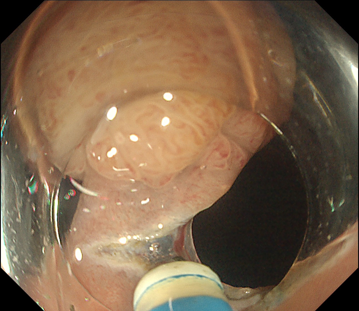
14. Final cut

Due to the lesion‘s large size and with the aid of gravity, ideal traction could be achieved during the entire procedure. This marks the final cut of the ESD.

Enhancement : A7
NBI Mode : NA
TXI Mode : NA
RDI Mode : NA
BAI-MAC : On

15. Completion of ESD

Lesion was resected en bloc successfully.

Enhancement : A7
NBI Mode : NA
TXI Mode : NA
RDI Mode : NA
BAI-MAC : On

Case Video

Overall Comment

This is a large rectal tumor (7 cm in diameter) detected by FIT screening and subsequent colonoscopy. Initially, it was examined using a high-definition colonoscope with magnification function. The lesion size exceeded half of the rectal circumference, and its macroscopic morphology was interpreted as 0-Is+IIa (LST-G-NM). According to a previous meta-analysis, lesions with such morphology carry a higher risk of submucosal invasion, estimated at around 10% (Bogie R et al. Endoscopy. 2018;50:263-282). While invasive cancer was reported to mainly occur in the large nodular part, a later study on a large ESD cohort revealed that invasive cancer may also be present in a multi-focal fashion and even in the non-nodular portion, justifying en bloc resection for such lesions (Yamada M et al. Endoscopy. 2016;48:456-64).

In this case, a focal JNET-2B capillary pattern was observed, indicative of advanced histology including high-grade dysplasia, carcinoma in situ, or even superficial invasive cancer. However, chromoendoscopy with crystal violet revealed a focal Kudo type Vi pit pattern without a demarcation line, indicating a non-invasive lesion and justifying endoluminal therapy.

ESD was performed using the Olympus DualKnife J (KD-655U) with a therapeutic colonoscope (PCF-H290TI), featuring an up angulation of 210° and a short bending section, facilitating the ESD procedure.

Histologically, this lesion was diagnosed as a tubulovillous adenoma with high-grade dysplasia, which was consistent with the area where the JNET 2B capillary pattern and Kudo type Vi pit pattern were observed.

* Specifications, design and accessories are subject to change without any notice or obligation on the part of the manufacturer

Keyword
Content Type