Colorectal Case 16

Dr. Serhii Polishchuk
Gastrointestinal endoscopist at LLC “GASTROCENTER” Olymed; Trainer at NGO “EndoAcademy”, Ukraine
Scope: CF-EZ1500DL
Organ: Colon (sigmoid colon)
Patient information: M, 80s, come for colonoscopy with bowel habit change
Medical history: Bowel habit change (constipation). No family history of CRC. No alcohol abuse, no smoking
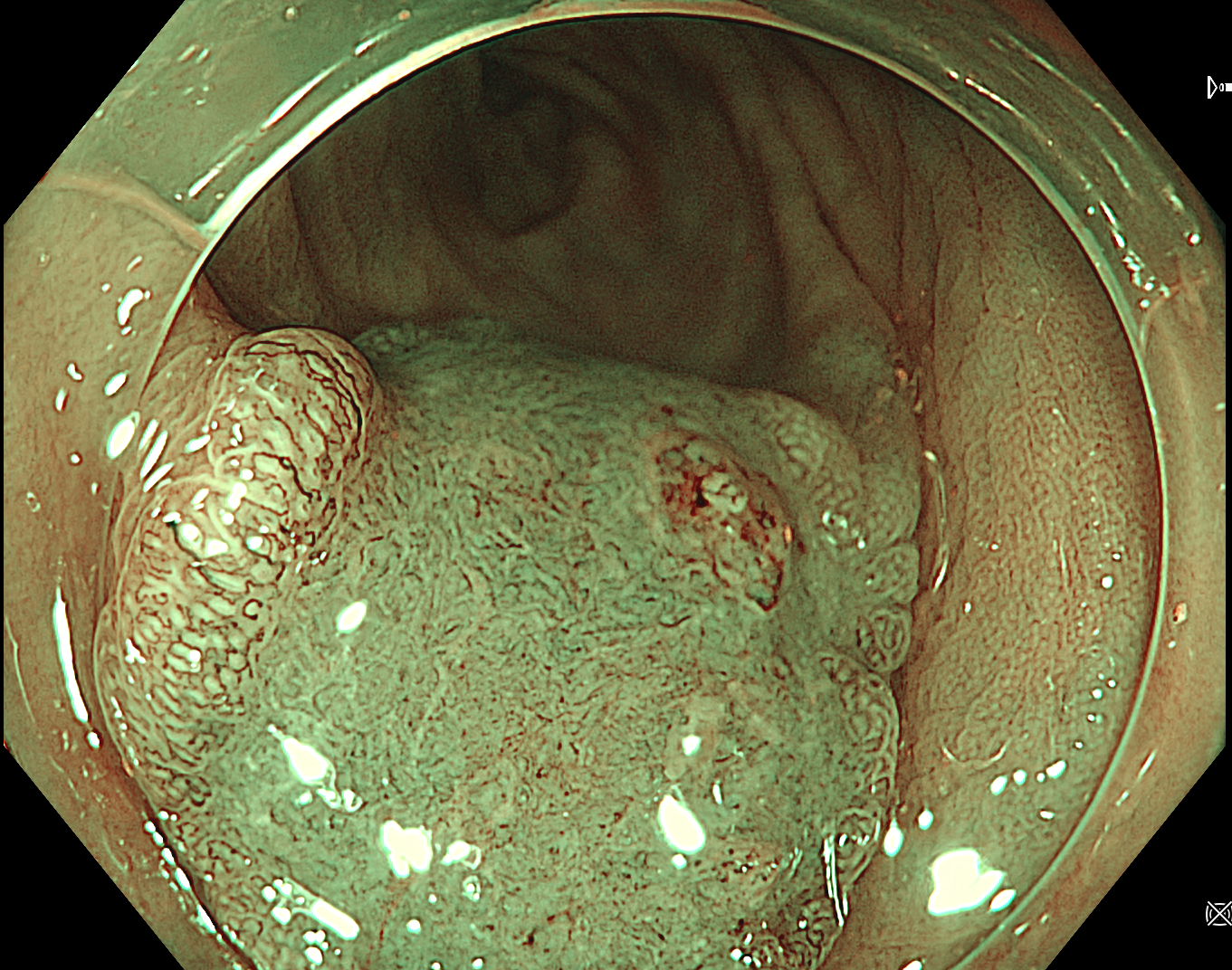
1. SM deep invasive cancer of colon
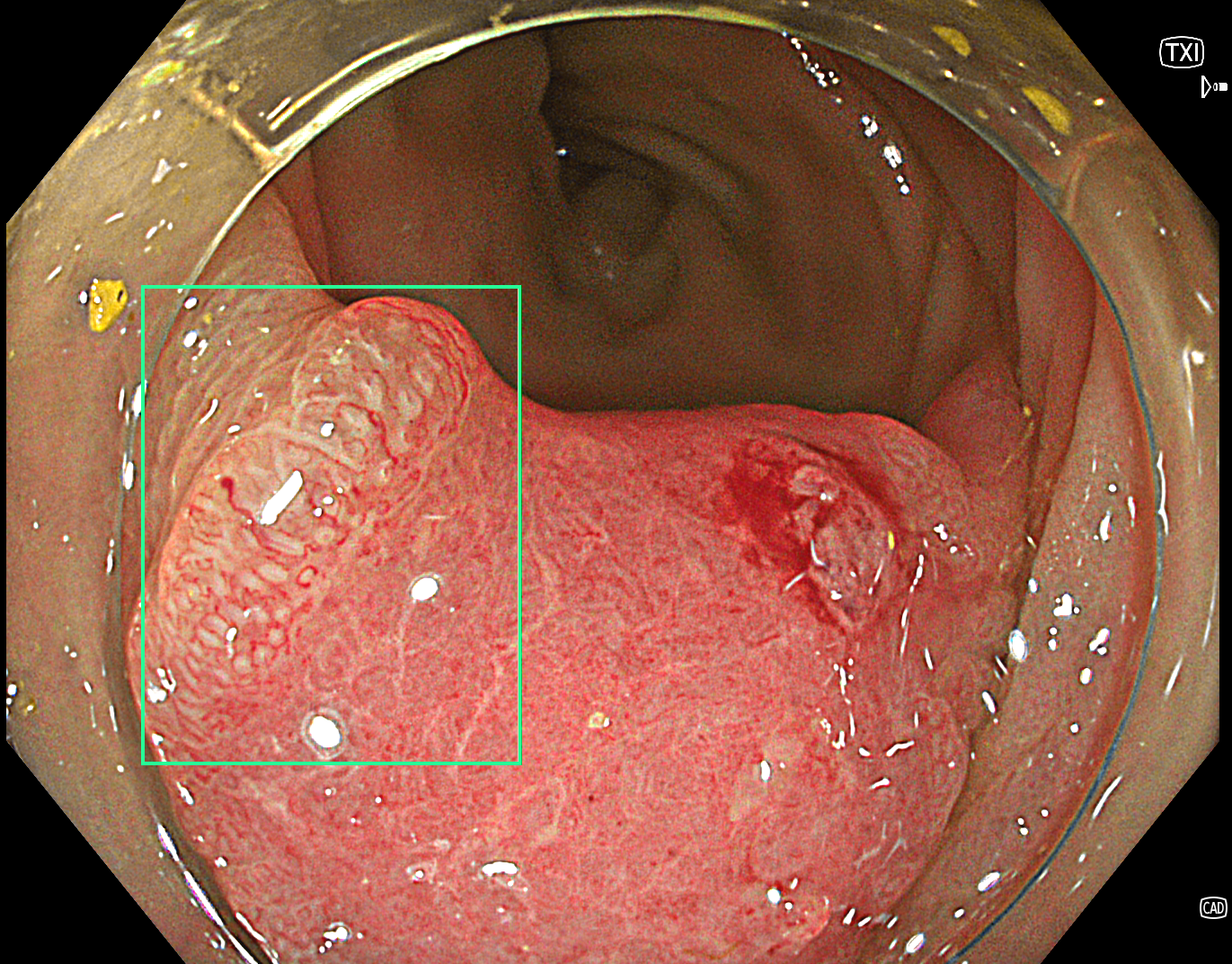
Texture and Color Enhancement Imaging (TXI): Mode 1
2. SM deep invasive cancer of colon
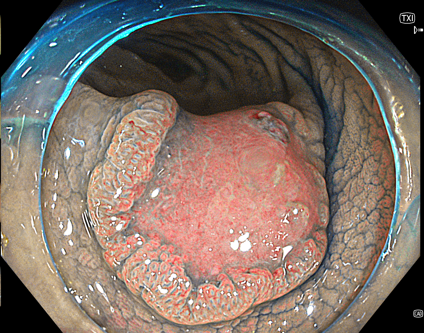
Texture and Color Enhancement Imaging (TXI): Mode 1
3. SM deep invasive cancer of colon
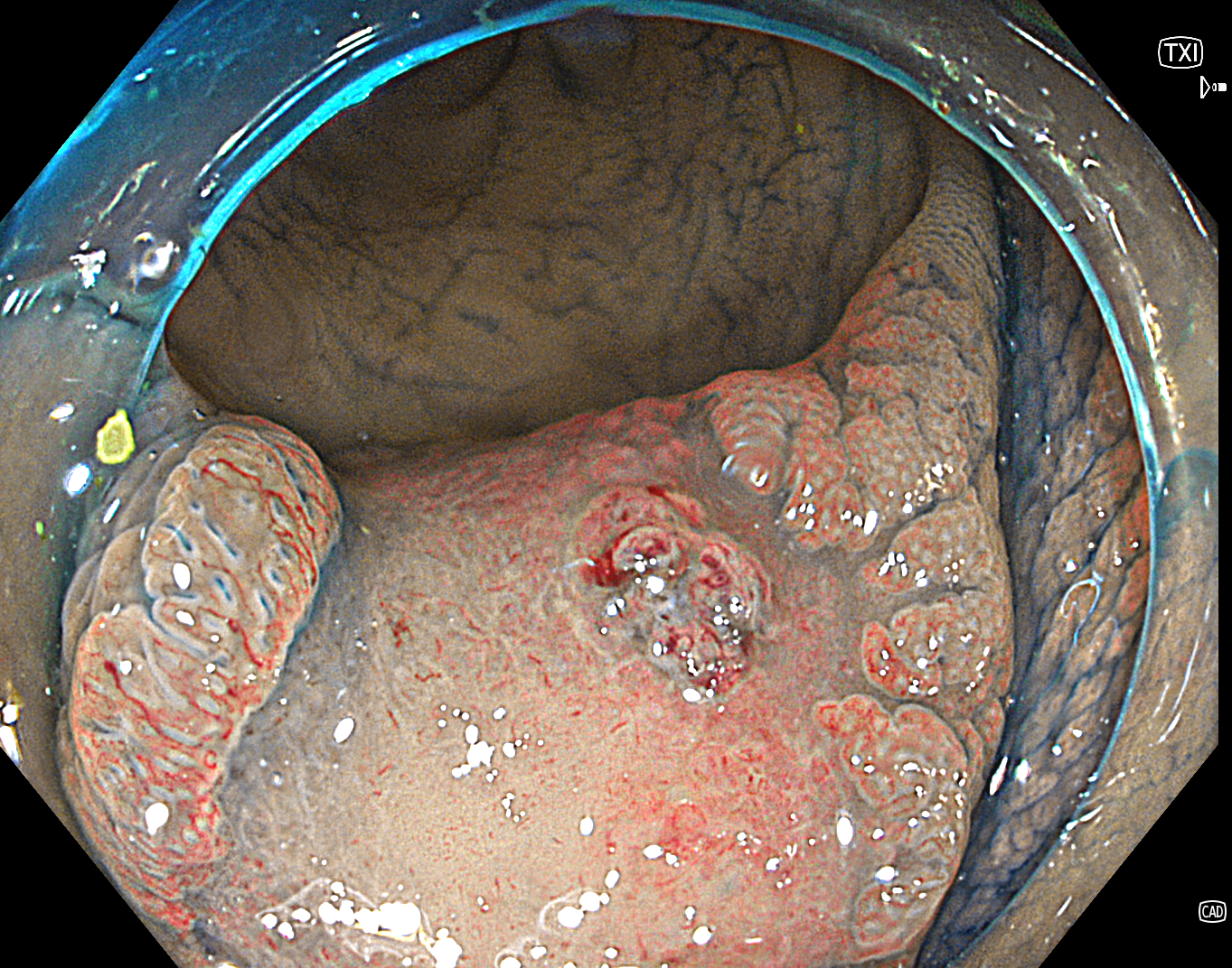
Texture and Color Enhancement Imaging (TXI): Mode 1
4. SM deep invasive cancer of colon
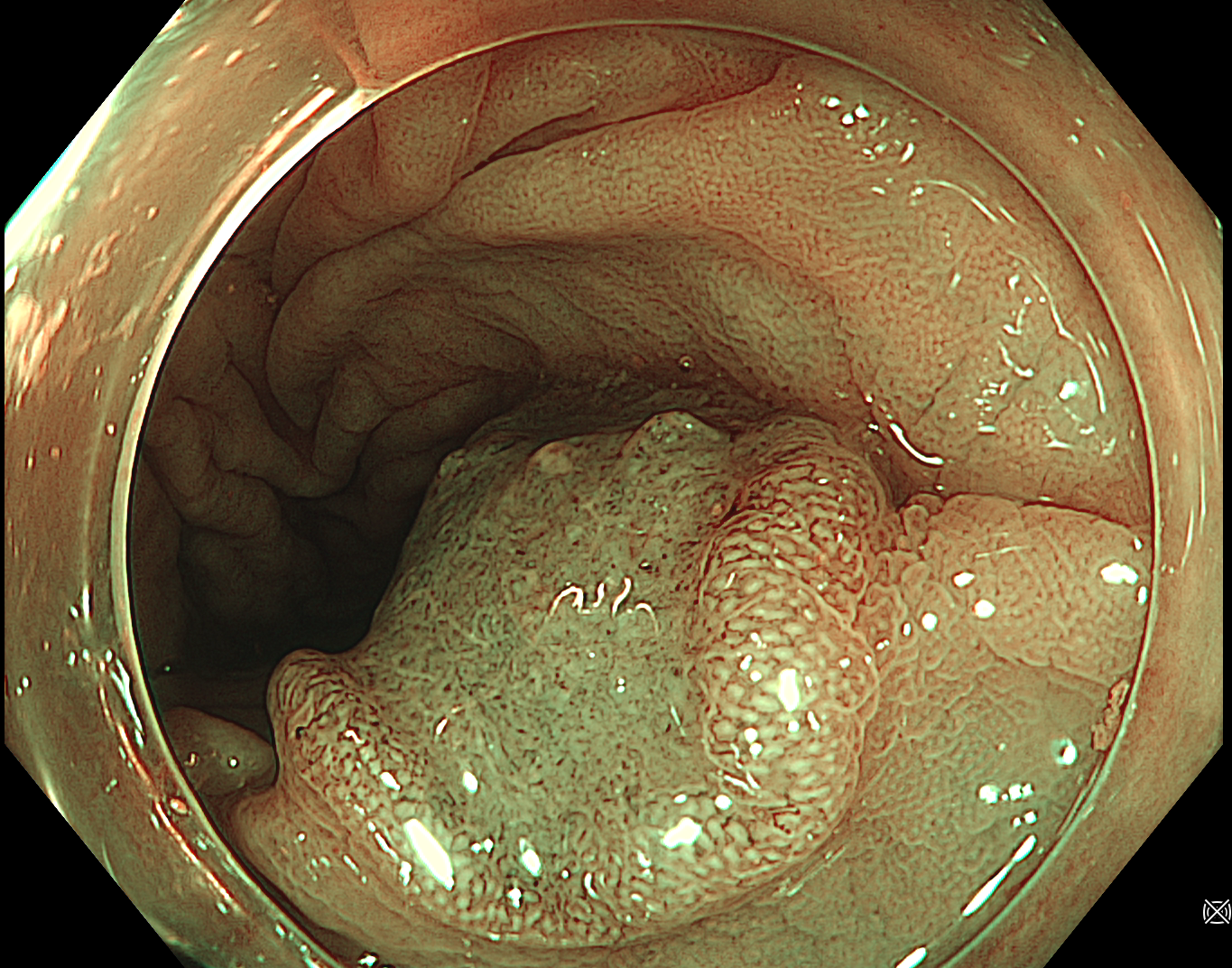
NBI Color Mode 3
Enhancement: A5
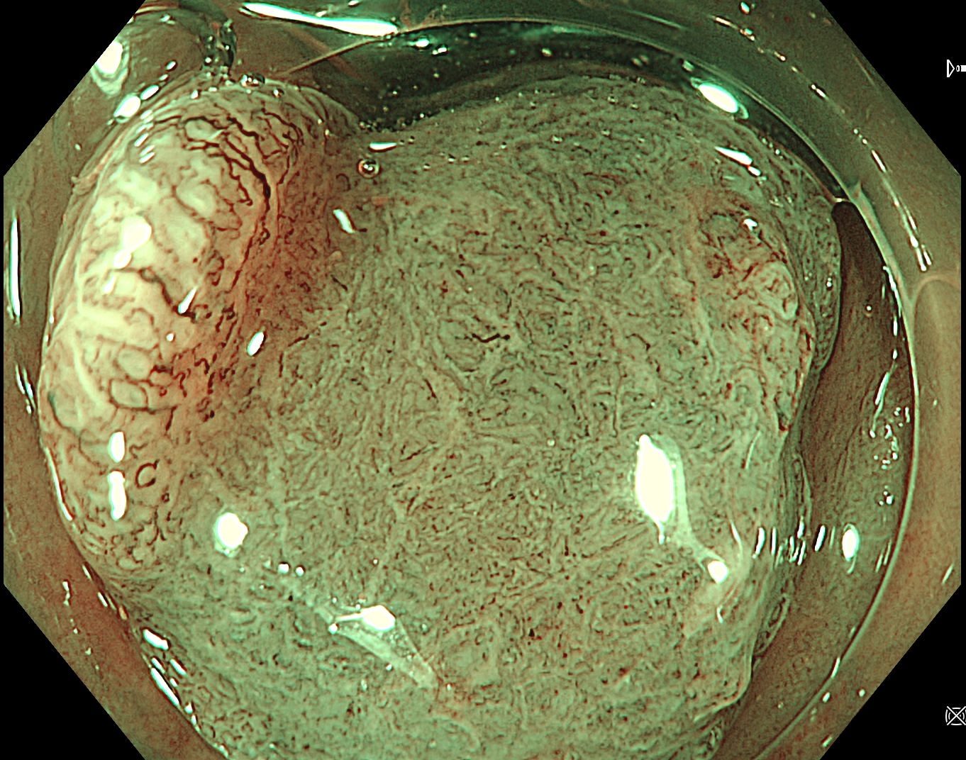
5. SM deep invasive cancer of colon
NBI Color Mode 3
Enhancement: A5
6. SM deep invasive cancer of colon
NBI Color Mode 3
Enhancement: A5
Case Video
SM deep invasive cancer of colon - JNET3
Overall Comment
The final optical diagnosis in this case was:
Location: sigmoid colon
Size: ~13mm
Morphology (Paris classification): 0-IIa+IIc
Pit pattern: Kudo classification IIIL+Vi+Vn
JNET classification: JNET3 (vessel pattern – loose vessel areas, interruption of thick vessels; amorphous areas)
Optical diagnosis: submucosal deep invasive cancer
Treatment: surgical resection
Pathology report: well differentiated adenocarcinoma of the sigmoid colon pT2N0R0LV1Pn0G1 (ICD-O code: 8140/3)
* Specifications, design and accessories are subject to change without any notice or obligation on the part of the manufacturer
Dr. Serhii Polishchuk Case 17: Tubular adenoma with LGD - JNET2A
Dr. Serhii Polishchuk
- Keyword
- Content Type


