Colorectal Case 12

Scope: CF-XZ1200
Patient information: M, 60s
Medical history: Previously fit and well; had FIT positive during screening
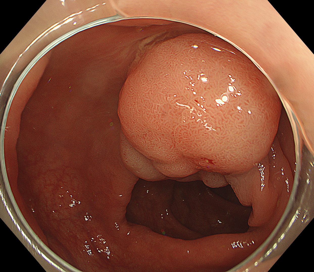
1. WLI observation
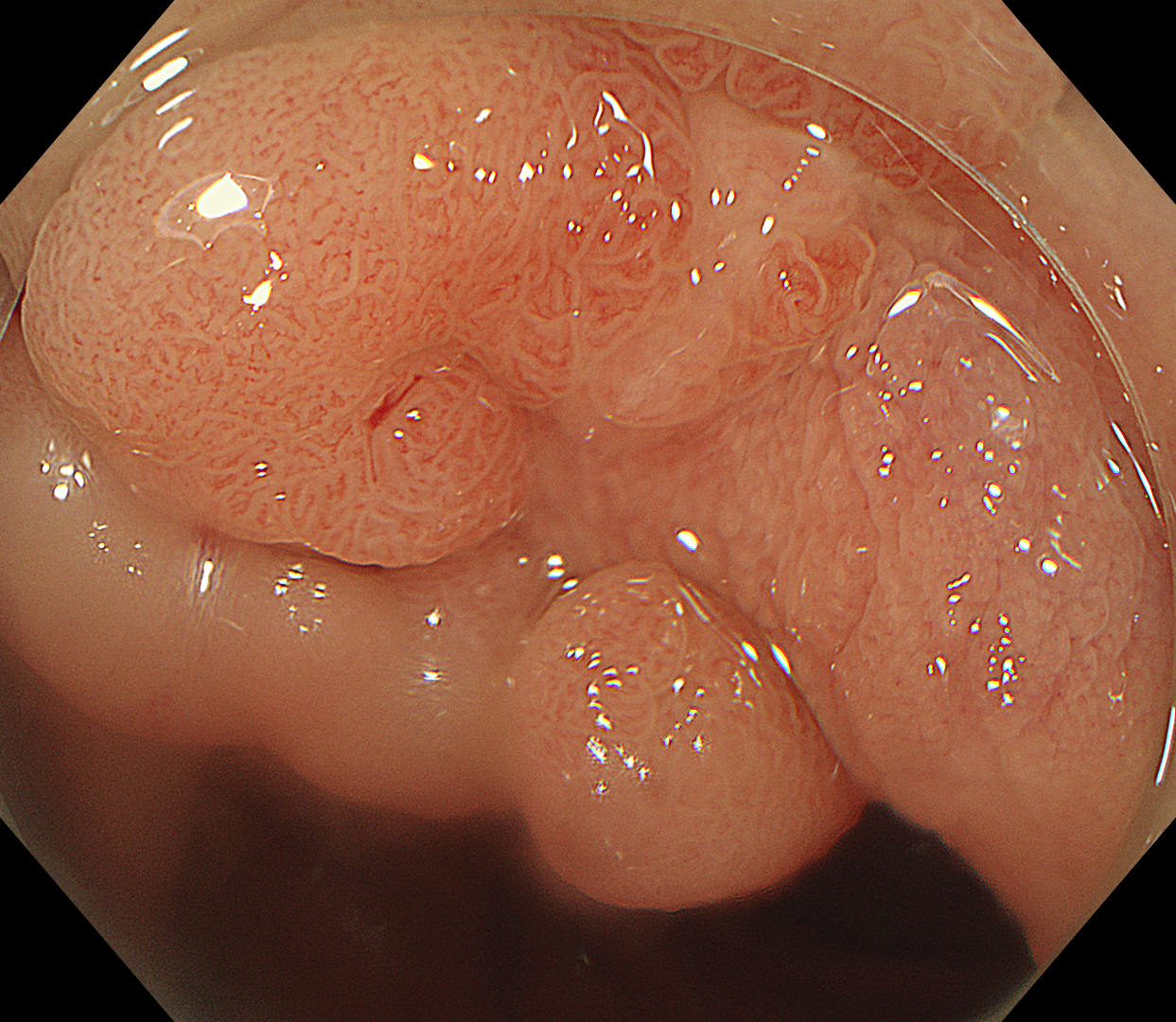
2. WLI with magnification observation
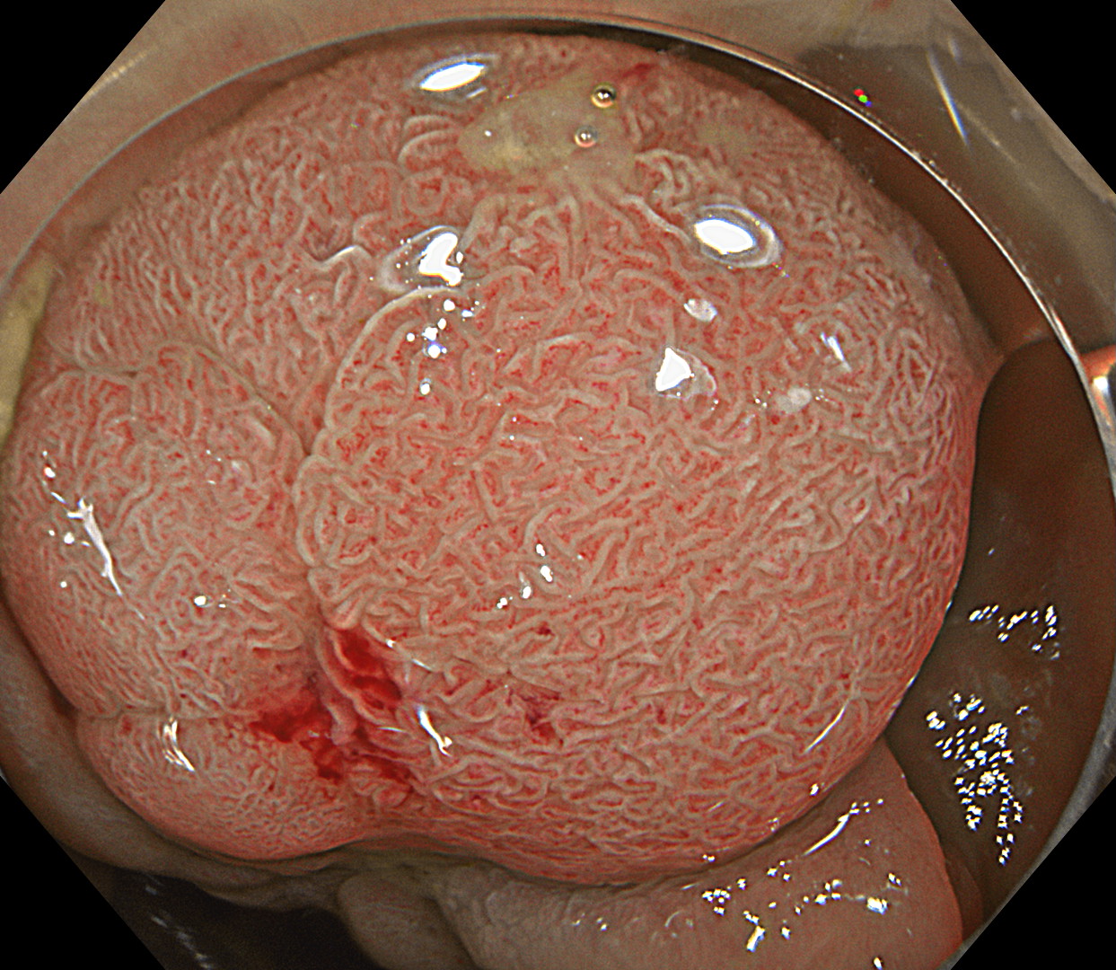
3. TXI Observation
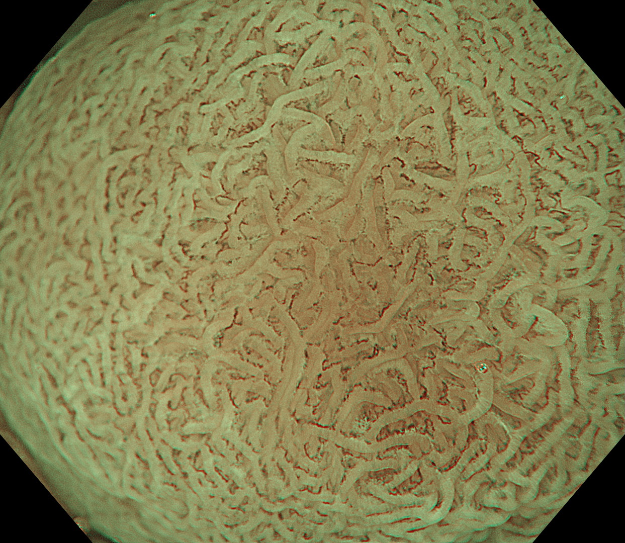
4. NBI Observation with optical magnification
Case Video
Overall Comment
EVIS X1 with CF-XZ1200 colonoscope provide excellent image quality over previous generations of endoscope system. The clarity and resolution of the endoscopic image are excellent, with the help of TXI function this will enhance the identification of lesions during examination. Once a lesion has been identified, the clear focused image contribute accurate detailed examination of morphology of the lesion. The excellent image quality extends to NBI mode, and together with optical magnification accurate characterisation of the lesion can be made. Ultimately this will help the endoscopist to make an accurate assessment of the lesion and subsequent treatment proposal.
* Specifications, design and accessories are subject to change without any notice or obligation on the part of the manufacturer
Dr. Supakij Khomvilai Case 13: Sessile Serrated Lesion (SSL) - JNET1
Dr. Serhii Polishchuk
- Content Type


