Colorectal Case 31

Prof. Naohisa Yahagi
Cancer Center, Keio University Hospital
Japan
Scope: PCF-H190I
Organ:Rectum
Patient information: M, 70s
Medical history: N/A
RDI™ technology is not intended to replace histopathological sampling as a means of diagnosis.
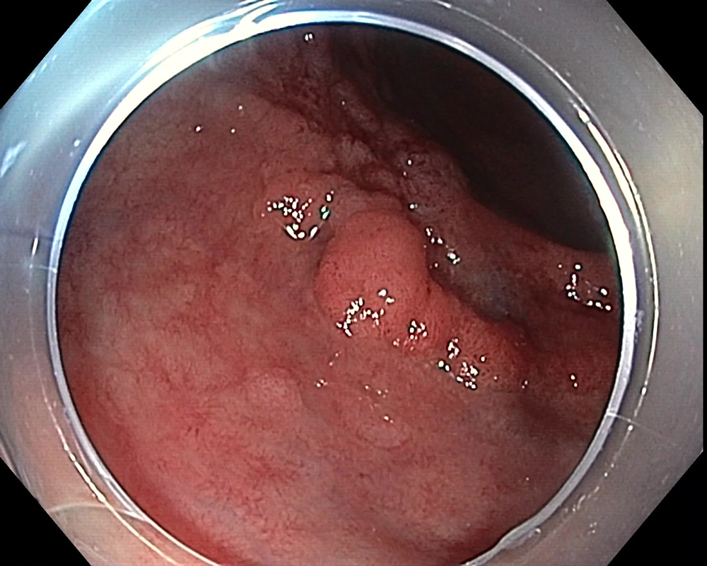
1. WLI Observation
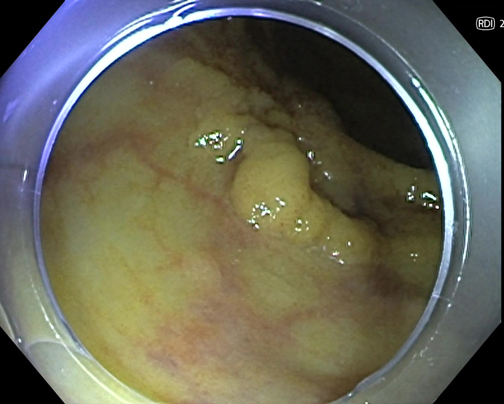
2. RDI Mode 2 observation
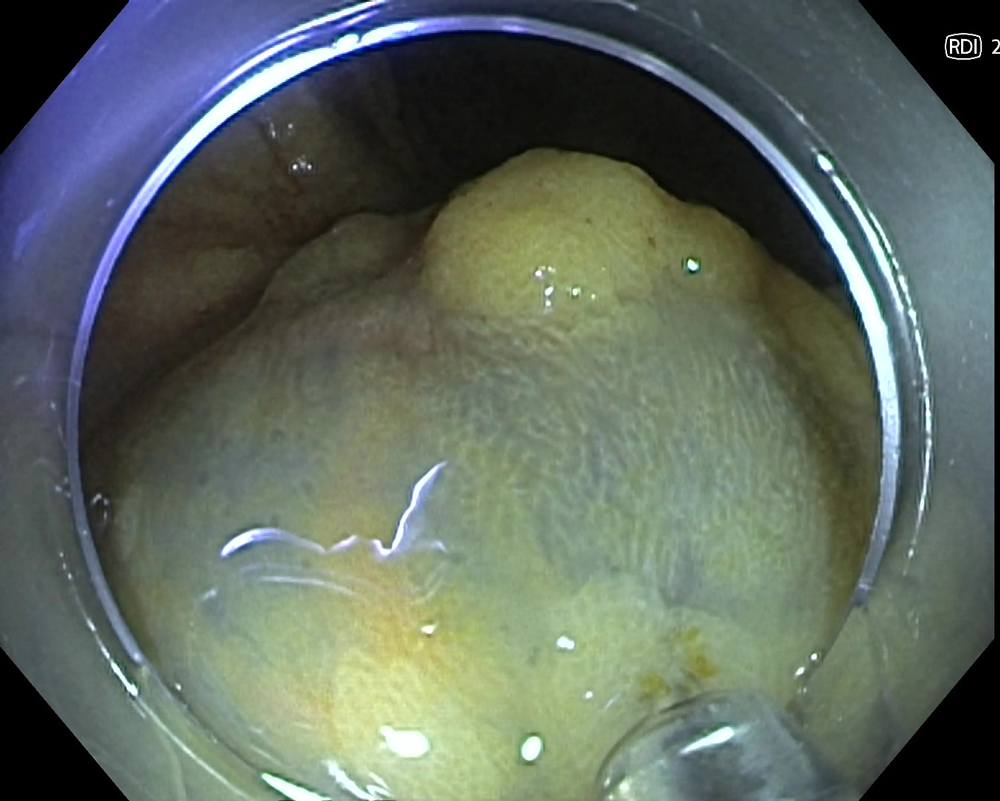
3. Injection under RDI Mode 2

4. Injection under RDI Mode 2
Case Video
5. Dissection
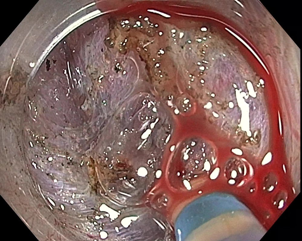
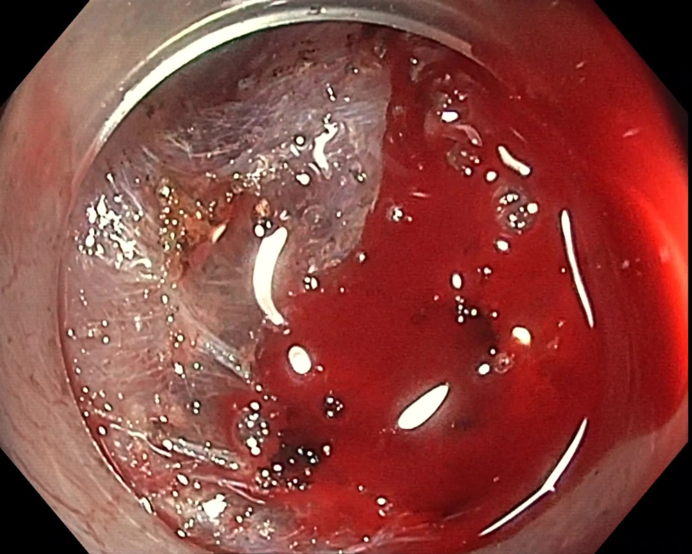
6. Bleeding
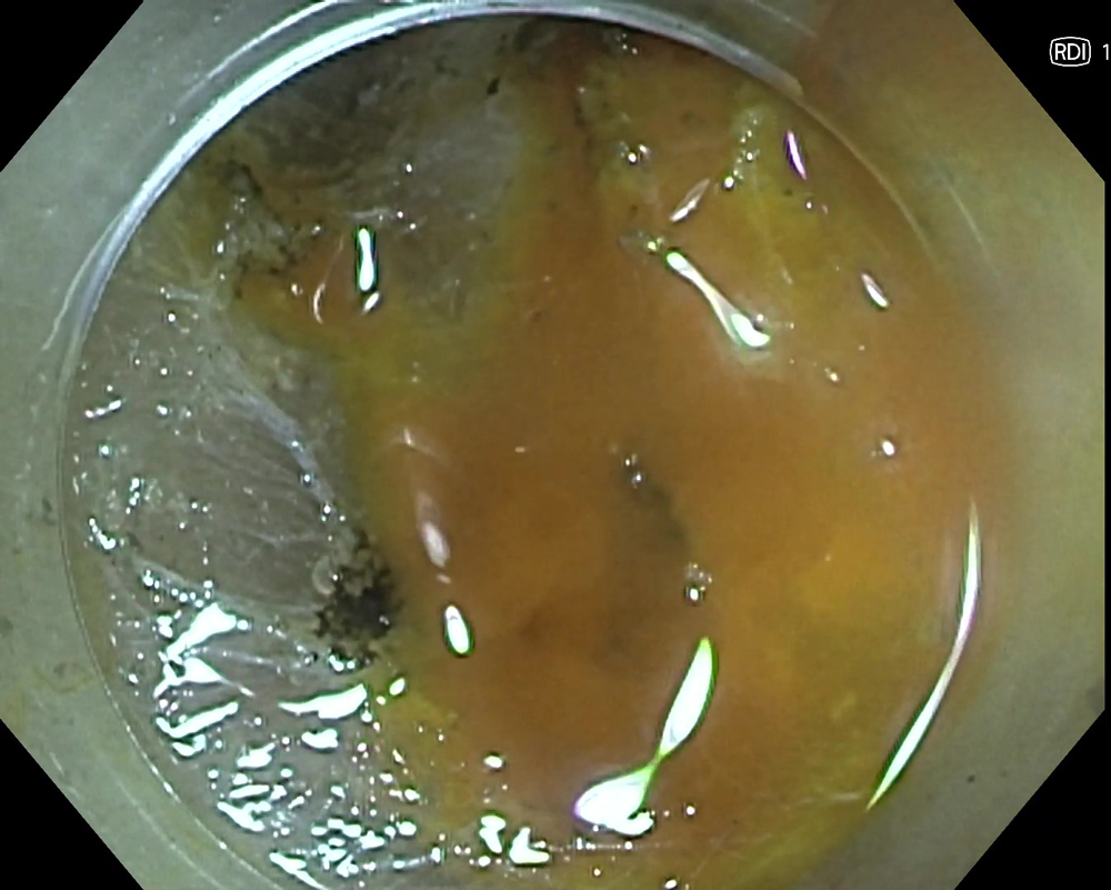
7. RDI Mode 1
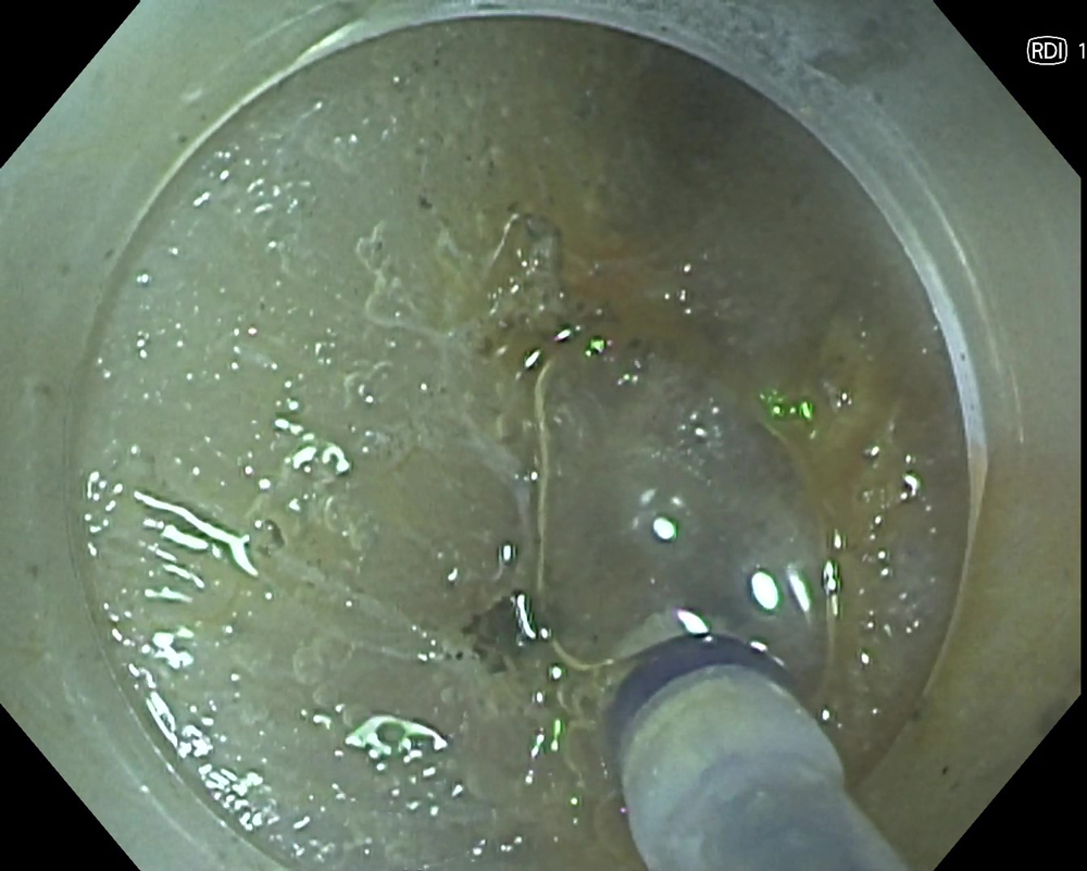
8. Hemostasis
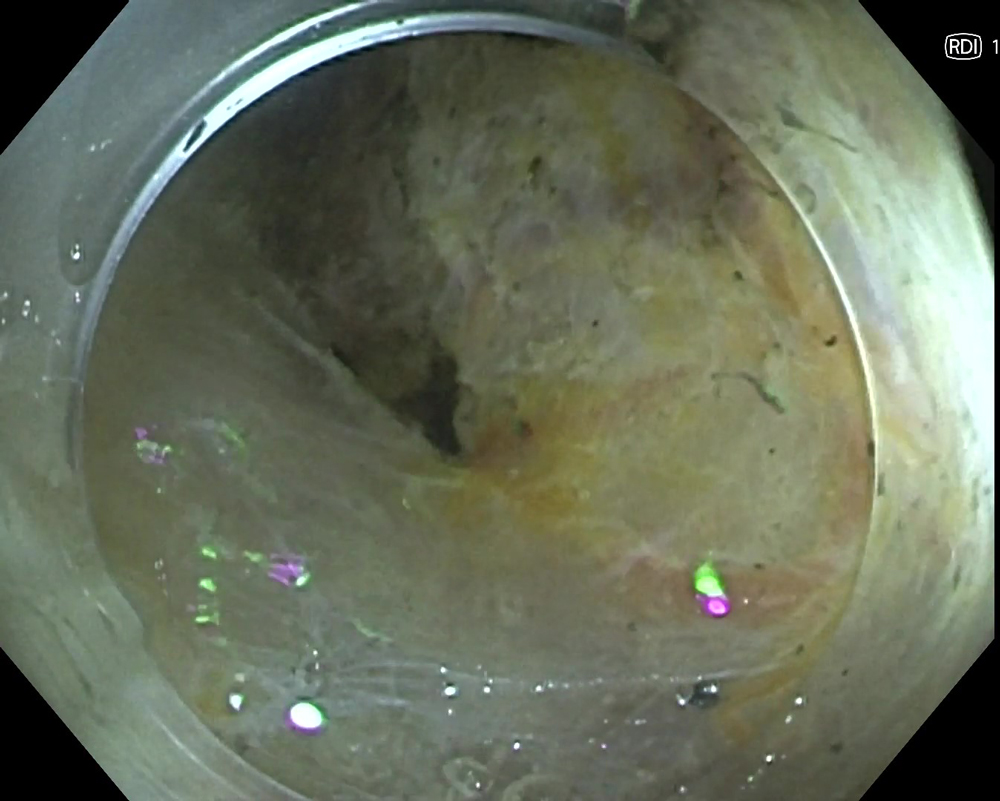
9. Completion
Case Video
Overall Comment
Although no studies are yet to verify the effectiveness of RDI technology, in my experience the use of RDI technology may help reduce hemostasis and treatment times. I have experienced the application of RDI technology to support hemostasis of gastrointestinal bleeding and diagnosis or treatment of esophageal varices in my medical center. Additionally, I have been experiencing the benefits of RDI technology use in cases outside of ESD.
I feel strong psychological stress when the entire monitor view turns red due to bleeding. I believe that visualizing hemorrhages in amber color instead of red decreases the sense of urgency and stress of operators towards bleeding, which allows operators to identify the bleeding point easily and perform hemostasis in a composed manner.
I expect that the efficiency and quality of endoscopic diagnosis and treatment are supported using the multiple functions of the EVIS X1™ endoscopy system, including RDI technology.
1) Data on file with Olympus (DC00489968)
2) Uraoka T, Igarashi M. “Development and clinical usefulness of a unique red dichromatic imaging technology in gastrointestinal endoscopy: A narrative review.” Therapeutic Advances in Gastroenterology. 2022;15.
* Specifications, design and accessories are subject to change without any notice or obligation on the part of the manufacturer


