Colorectal Case 33

Prof. Dr. Fatih Aslan
Koc University Hospital
Istanbul, Turkey
Scope: CF-XZ1200
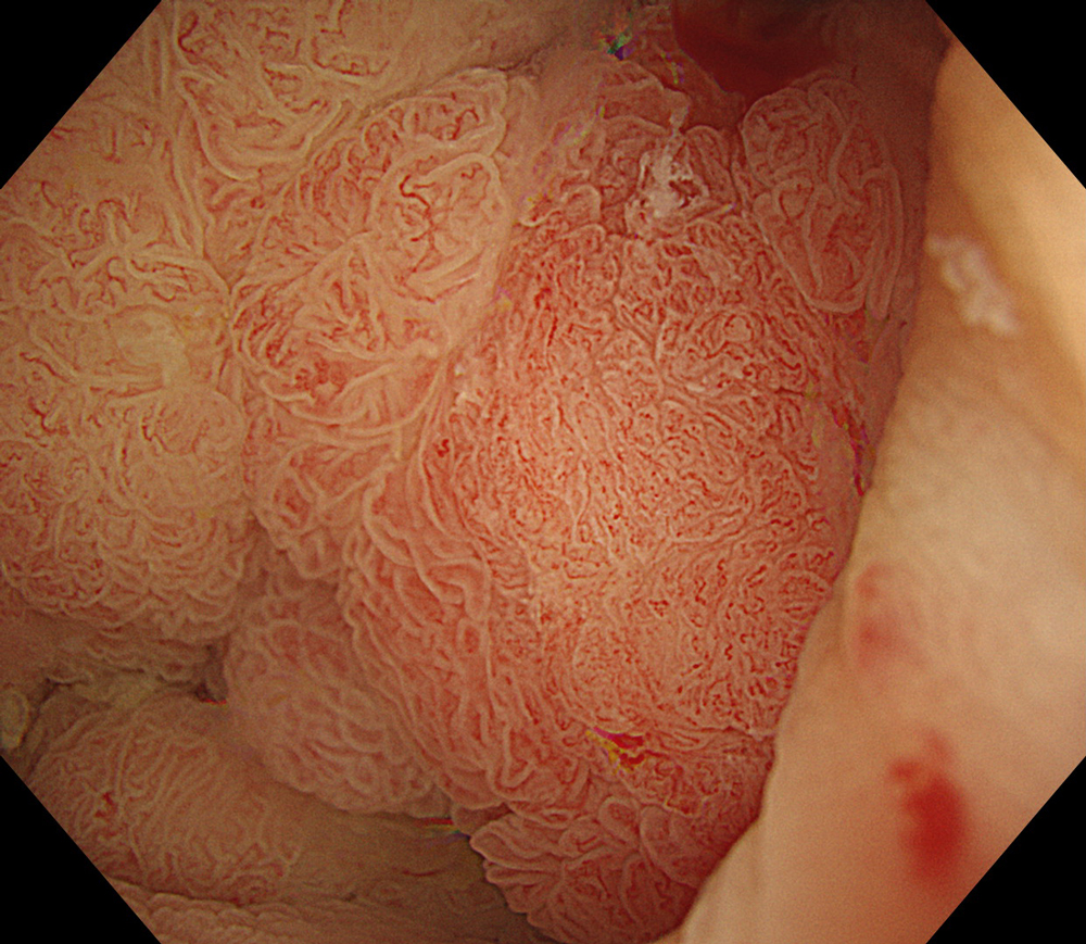
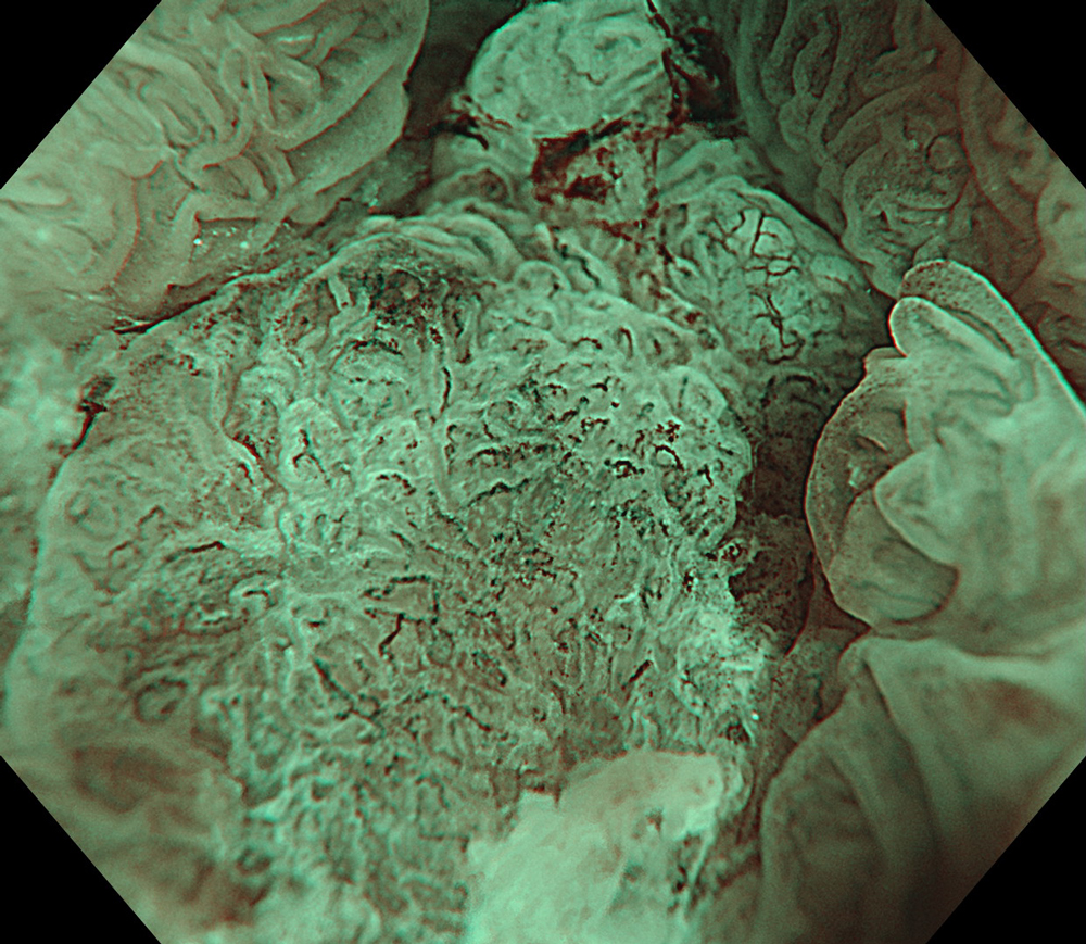
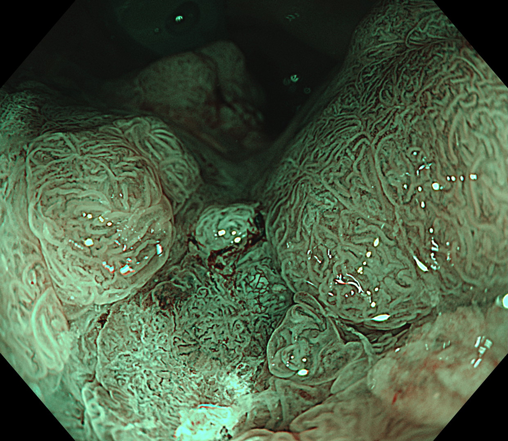
Case: LST-GM, invasive cancer
Organ: Rectum
1. WLI
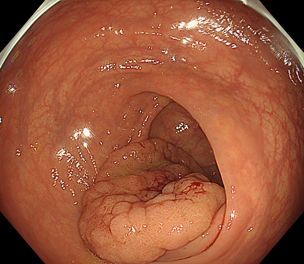
#WLI #A8 structure enhancement #Auto Iris
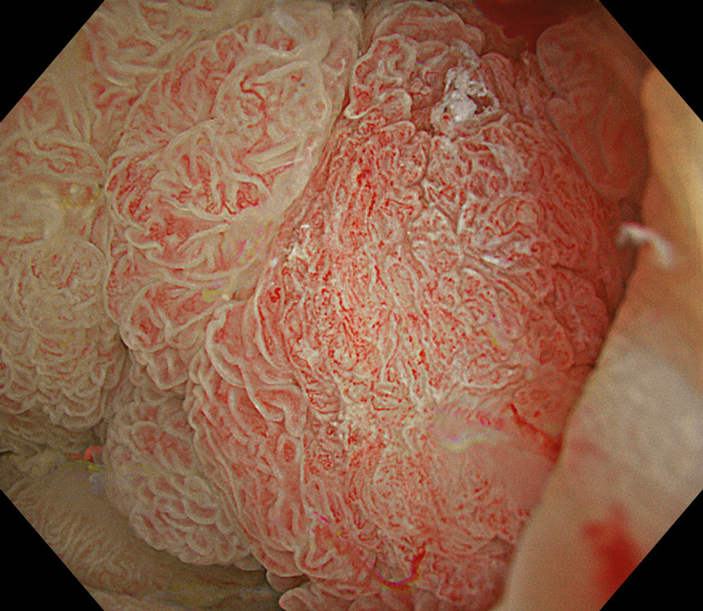
2. WLI-magnification
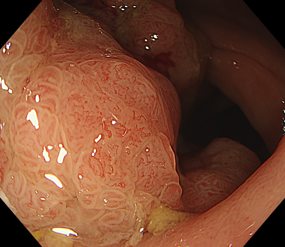
#WLI #A8 structure enhancement #optic magnification
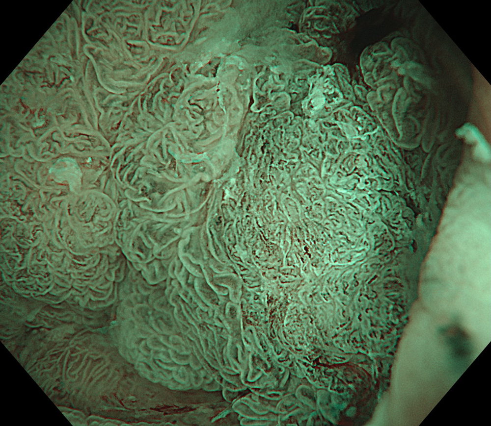
3. NBI-magnification
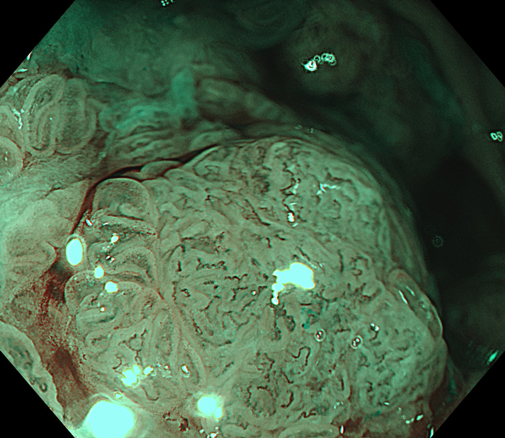
#NBI #A8 structure enhancement #optic magnification
4. NBI-optic magnification
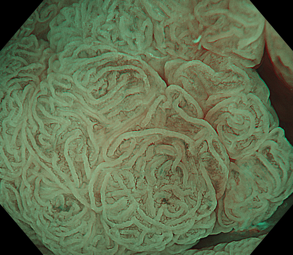
#NBI #A8 structure enhancement #optic magnification
5. WLI
#WLI #A8 structure enhancement #Auto Iris
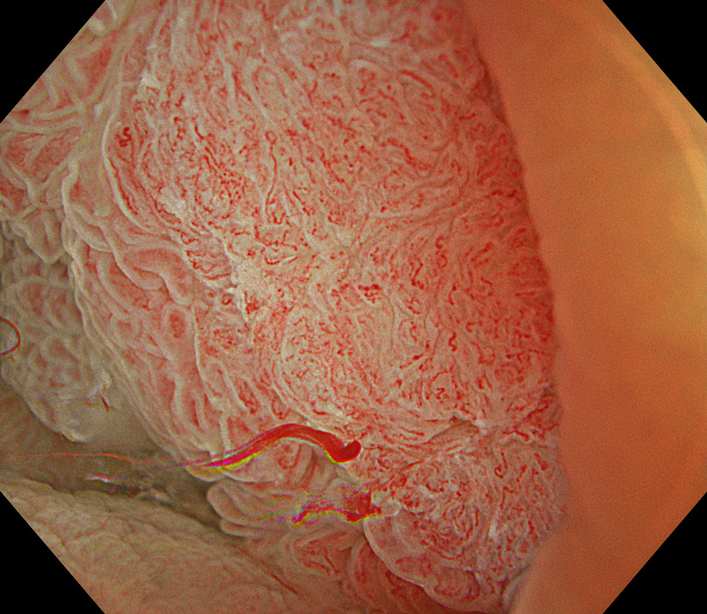
6. TXI
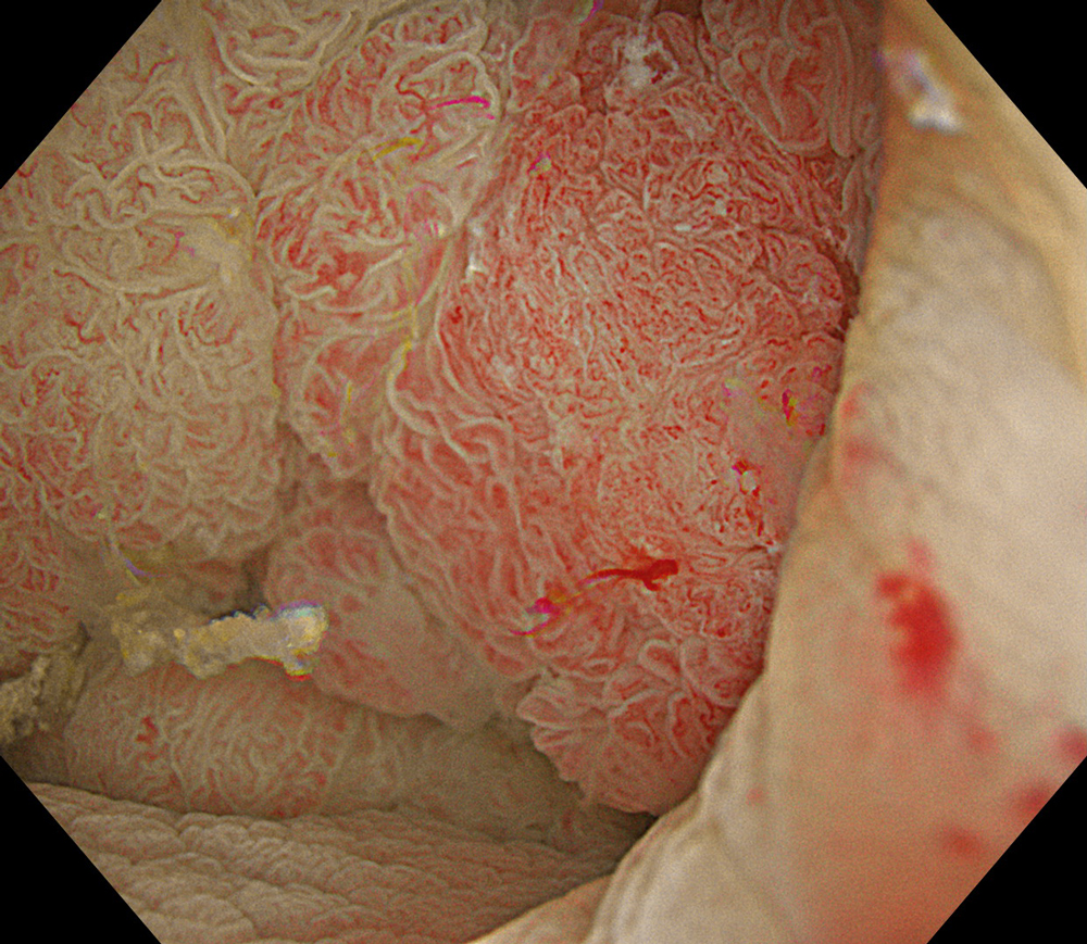
#TXI #A8 structure enhancement #Auto Iris
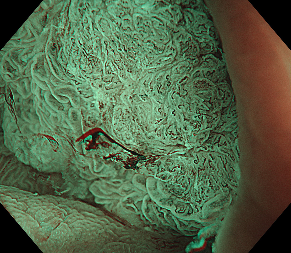
7. NBI
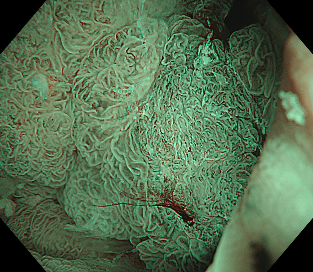
#NBI #A8 structure enhancement #optic magnification
8. TXI
#TXI #A8 structure enhancement #optic magnification
9. NBI
#NBI #A8 structure enhancement #optic magnification
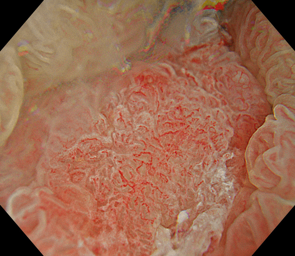
10. TXI
#TXI #A8 structure enhancement #optic magnification
11. NBI
#NBI #A8 structure enhancement #optic magnification
12. WLI-magnification

#WLI #A8 structure enhancement #optic magnification
13. TXI-magnification
#TXI #A8 structure enhancement #optic magnification
14. NBI-magnification
#NBI #A8 structure enhancement #optic magnification
15. NBI
#NBI #A8 structure enhancement
Case Video
High-definition imaging with the Olympus CF-XZ1200 endoscope and its optical magnification feature allowed for meticulous inspection of the lesion. The integration of TXI and NBI modes, in conjunction with underwater magnified observation, proved particularly valuable in delineating suspicious zones and assessing potential submucosal invasion, as clearly seen in the video documentation.
Overall Comment
A 66-year-old female patient presented with rectal bleeding. Colonoscopic examination revealed a granular mixed-type laterally spreading tumor-mixed type measuring approximately 8 cm in diameter. Random biopsies were reported as tubular adenoma with low-grade dysplasia. The patient was referred to our center for endoscopic resection.
The lesion was evaluated using an Olympus CF-XZ1200 endoscope equipped with optical magnification. With the use of TXI and NBI modes, the entire lesion surface was examined in detail. As demonstrated in the accompanying images and video, these imaging modes—combined with underwater observation and optical magnification—enabled a thorough assessment of suspicious areas, particularly with respect to potential submucosal invasion.
In this case, targeted biopsies taken from areas suspicious for malignancy were reported as invasive carcinoma. Radiological evaluation revealed malignant lymph nodes in the mesorectum, and the lesion was observed to have invaded the muscularis propria in certain regions.
Next-generation magnifying endoscopes, together with advanced imaging technologies such as TXI and NBI, significantly enhance the ability to perform targeted biopsies and improve diagnostic accuracy. Moreover, accurate diagnosis and staging through these modalities facilitate the selection of the most appropriate therapeutic approach.
* Specifications, design and accessories are subject to change without any notice or obligation on the part of the manufacturer.


