- Home
- Search: NBI
Search: NBI
-
 Add to View
 EVIS X1 Atlas - Imaging AtlasCase 10: Post-eradication gastric cancer screening Dr. Kunihisa Uchita
 -
 Add to View
Gastroenterology - Imaging AtlasCases 2: Longitudinal ulcer scars Dr. Kazuo Ohtsuka
Add to View
Gastroenterology - Imaging AtlasCases 2: Longitudinal ulcer scars Dr. Kazuo Ohtsuka -
 Add to View
Colorectal - Imaging AtlasCase 29: Multiple lesions in ascending colon Prof. Dr. Hu Xiao
Add to View
Colorectal - Imaging AtlasCase 29: Multiple lesions in ascending colon Prof. Dr. Hu Xiao -
Add to View
EVIS X1 Atlas - Imaging AtlasCase 29: Multiple lesions in ascending colon Prof. Dr. Hu Xiao
-
Add to View
Colorectal - Imaging AtlasCase 29: Multiple lesions in ascending colon Prof. Dr. Hu Xiao
-
Add to View
Colorectal - Imaging AtlasCase 31: Multiple lesions in ascending colon Prof. Dr. Hu Xiao
-
Add to View
EVIS X1™ Atlas - Imaging AtlasCase 31: Multiple lesions in ascending colon Prof. Dr. Hu Xiao
-
Add to View
EVIS X1 Atlas - Imaging AtlasCase 30: Multiple lesions in ascending colon Prof. Dr. Hu Xiao
-
Add to View
EVIS X1 Atlas - Imaging AtlasCase 8: Early stomach cancer Prof. Dr. Liu Zhiguo
-
Add to View
Gastroenterology - Imaging AtlasCase 8: Early stomach cancer Prof. Dr. Liu Zhiguo
-
Add to View
Gastroenterology - Imaging AtlasCase 8: Early stomach cancer (M cancer) Prof. Dr. Liu Zhiguo
-
 Add to View
Gastroenterology - Imaging AtlasCase 10: Barrett neoplasia Dr. Roos Pouw
Add to View
Gastroenterology - Imaging AtlasCase 10: Barrett neoplasia Dr. Roos Pouw -
Add to View
Gastroenterology - Imaging AtlasCase 10: Barrett neoplasia Dr. Roos Pouw
-
Add to View
Gastroenterology - Imaging AtlasCase 10: Barrett neoplasia Dr. Roos Pouw
-
Add to View
EVIS X1 Atlas - Imaging AtlasCase 10: Barrett neoplasia Dr. Roos Pouw
-
Add to View
EVIS X1 Atlas - Imaging AtlasCase 10: Barrett neoplasia Dr. Roos Pouw
-
Add to View
EVIS X1™ Atlas - Imaging AtlasCase 7: Barrett Esophagus-Adenocarcinoma Prof. Dr. Fatih Aslan
-
Add to View
Gastroenterology - Imaging AtlasCase 11: Barrett Esophagus-Adenocarcinoma Prof. Dr. Fatih Aslan
-
Add to View
Gastroenterology - Imaging AtlasCase 11: Barrett Esophagus-Adenocarcinoma Prof. Dr. Fatih Aslan
-
Add to View
Esophageal - Imaging AtlasCase 11: Barrett Esophagus-Adenocarcinoma Prof. Dr. Fatih Aslan
-
Add to View
Esophageal - Imaging AtlasCase 10: Barrett neoplasia Dr. Roos Pouw
-
Add to View
Gastroenterology - Imaging AtlasCase 9: Barrett esophagus inspection Dr. Roos Pouw
-
Add to View
Gastroenterology - Imaging AtlasCase 9: Barrett esophagus inspection Dr. Roos Pouw
-
Add to View
Gastroenterology - Imaging AtlasCase 9: Barrett esophagus inspection Dr. Roos Pouw
-
Add to View
EVIS X1 Atlas - Imaging AtlasCase 9: Barrett esophagus inspection Dr. Roos Pouw
-
Add to View
EVIS X1 Atlas - Imaging AtlasCase 9: Barrett esophagus inspection Dr. Roos Pouw
-
Add to View
Esophageal - Imaging AtlasCase 9: Barrett esophagus inspection Dr. Roos Pouw
-
Add to View
Gastroenterology - Imaging AtlasCase 8: Eosinophilic esophagitis Dr. Khanh Do-Cong Pham
-
Add to View
Gastroenterology - Imaging AtlasCase 8: Eosinophilic esophagitis Dr. Khanh Do-Cong Pham
-
Add to View
EVIS X1 Atlas - Imaging AtlasCase 8: Eosinophilic esophagitis Dr. Khanh Do-Cong Pham
-
Add to View
EVIS X1 Atlas - Imaging AtlasCase 8: Eosinophilic esophagitis Dr. Khanh Do-Cong Pham
-
Add to View
Gastroenterology - Imaging AtlasCase 7: Two gastric neuroendocrine tumors Dr. Khanh Do-Cong Pham
-
Add to View
Gastroenterology - Imaging AtlasCase 7: Two gastric neuroendocrine tumors Dr. Khanh Do-Cong Pham
-
Add to View
Gastroenterology - Imaging AtlasCase 7: Two gastric neuroendocrine tumors Dr. Khanh Do-Cong Pham
-
Add to View
EVIS X1 Atlas - Imaging AtlasCase 7: Two gastric neuroendocrine tumors Dr. Khanh Do-Cong Pham
-
Add to View
EVIS X1 Atlas - Imaging AtlasCase 6: Two gastric neuroendocrine tumors Dr. Khanh Do-Cong Pham
-
Add to View
Colorectal - Imaging AtlasCase 29: Is+IIa (LST-G) lesion Dr. Hiroaki Ikematsu
-
Add to View
Colorectal - Imaging AtlasCase 30: Is+IIa (LST-G) lesion Dr. Hiroaki Ikematsu
-
Add to View
EVIS X1™ Atlas - Imaging AtlasCase 33: LST-GM, invasive cancer Prof. Dr. Fatih Aslan
-
Add to View
Colorectal - Imaging AtlasCases 33: LST-GM, invasive cancer Prof. Dr. Fatih Aslan
-
Add to View
Colorectal - Imaging AtlasCase 30: LST-GM, invasive cancer Prof. Dr. Fatih Aslan
-
Add to View
Colorectal - Imaging AtlasCase 31: LST-GM, invasive cancer Prof. Dr. Fatih Aslan
-
Add to View
- Imaging AtlasCases 34: A 30-mm non-granular laterally spreading tumor (LST-NG-FE) Prof. Yunho Jung
-
Add to View
EVIS X1 Atlas - Imaging AtlasCases 33: LST-GM, invasive cancer Prof. Dr. Fatih Aslan
-
Add to View
Gastroenterology - Imaging AtlasCase 6: Early gastric cancer - from screening to detailed examination Dr. Hisashi Doyama
-
Add to View
EVIS X1 Atlas - Imaging AtlasCase 6: Early gastric cancer - from screening to detailed examination Dr. Hisashi Doyama
-
Add to View
Gastric - Imaging AtlasCase 6: Early gastric cancer - from screening to detailed examination Dr. Hisashi Doyama
-
 Add to View
Colonoscopy - ReferenceEVIS X1 Endoscopist Interview
Add to View
Colonoscopy - ReferenceEVIS X1 Endoscopist Interview -
 Add to View
EVIS X1 Atlas - Imaging AtlasCase 11: Early gastric cancer Takashi Kawai, MD, PhD
Add to View
EVIS X1 Atlas - Imaging AtlasCase 11: Early gastric cancer Takashi Kawai, MD, PhD -
Add to View
Gastroenterology - Imaging AtlasCase 9: Early gastric cancer (tub2>tub1, 0-IIa+IIc, pT1b2(SM2), UL(-), ly0, v0) Takashi Kawai, MD, PhD
-
Add to View
EVIS X1 Atlas - Imaging AtlasCase 12: Early gastric cancer Takashi Kawai, MD, PhD
-
Add to View
Gastroenterology - Imaging AtlasCase 10: Early gastric cancer (por2>sig>tub2, 0-IIc, pT1a(M), UL(-), ly0, v0) Takashi Kawai, MD, PhD
-
Add to View
EVIS X1 Atlas - Imaging AtlasCase 13: Regular arrangement of collecting venules Takashi Kawai, MD, PhD
-
Add to View
Gastroenterology - Imaging AtlasCase 11: Regular arrangement of collecting venules (RAC) Takashi Kawai, MD, PhD
-
Add to View
EVIS X1 Atlas - Imaging AtlasCase 7: Superficial esophageal cancer Haruhiro Inoue, MD, PhD
-
Add to View
Gastroenterology - Imaging AtlasCase 7: Superficial esophageal cancer Haruhiro Inoue, MD, PhD
-
Add to View
Gastroenterology - Imaging AtlasCase 7: Superficial esophageal cancer Haruhiro Inoue, MD, PhD
-
Add to View
Esophageal - Imaging AtlasCase 7: Superficial esophageal cancer Haruhiro Inoue, MD, PhD
-
 Add to View
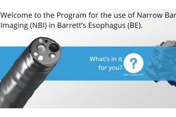
Upper GI Endoscopy - E-learningNBI Barrett's esophagus
Add to View
Upper GI Endoscopy - E-learningNBI Barrett's esophagus -
Add to View
Upper GI Endoscopy - E-learningNBI™ technology - Barrett's Esophagus
-
 Add to View
Colonoscopy - Technology InformationX1 Elevating the Standard of Endoscopy
Add to View
Colonoscopy - Technology InformationX1 Elevating the Standard of Endoscopy -
Add to View
Upper GI Endoscopy - Technology InformationX1 Elevating the Standard of Endoscopy
-
 Add to View
Existing Evidence Map- Pulmonology - Reference1: Navigation
Add to View
Existing Evidence Map- Pulmonology - Reference1: Navigation -
Add to View
EVIS X1 Atlas - Imaging AtlasCase 11: Colonic polyp (Tubular adenoma) Dr. Supakij Khomvilai
-
Add to View
Colorectal - Imaging AtlasCase 10: Colonic polyp (Tubular adenoma) Dr. Supakij Khomvilai
-
Add to View
Colorectal - Imaging AtlasCase 11: Colonic polyp (Tubular adenoma) Dr. Supakij Khomvilai
-
Add to View
Colorectal - Imaging AtlasCase 11: Colonic polyp (Tubular adenoma) Dr. Supakij Khomvilai
-
Add to View
Colorectal - Imaging AtlasCase 11: Colonic polyp (Tubular adenoma) Dr. Supakij Khomvilai
-
Add to View
EVIS X1™ Atlas - Imaging AtlasCase 11: Colonic polyp (Tubular adenoma) Dr. Supakij Khomvilai
-
Add to View
EVIS X1 Atlas - Imaging AtlasCase 11: Colonic polyp (Tubular adenoma) Dr. Supakij Khomvilai
-
Add to View
EVIS X1 Atlas - Imaging AtlasCase 8: Colonic polyp (Tubular adenom) Dr. Supakij Khomvilai
-
Add to View
Colorectal - Imaging AtlasCase 7: Colonic polyp (Tubular adenom) Dr. Supakij Khomvilai
-
Add to View
Colorectal - Imaging AtlasCase 8: Colonic polyp (Tubular adenom) Dr. Supakij Khomvilai
-
Add to View
Colorectal - Imaging AtlasCase 8: Colonic polyp (Tubular adenoma) Dr. Supakij Khomvilai
-
Add to View
EVIS X1™ Atlas - Imaging AtlasCase 8: Colonic polyp (Tubular adenom) Dr. Supakij Khomvilai
-
Add to View
EVIS X1 Atlas - Imaging AtlasCase 8: Colonic polyp (Tubular adenom) Dr. Supakij Khomvilai
-
Add to View
EVIS X1 Atlas - Imaging AtlasCase 9: Colonic polyp (Sessile Serrated Lesion) Dr. Supakij Khomvilai
-
Add to View
Colorectal - Imaging AtlasCase 8: Colonic polyp (Sessile Serrated Lesion) Dr. Supakij Khomvilai
-
Add to View
Colorectal - Imaging AtlasCase 9: Colonic polyp (Sessile Serrated Lesion) Dr. Supakij Khomvilai
-
Add to View
Colorectal - Imaging AtlasCase 9: Colonic polyp (Sessile Serrated Lesion) Dr. Supakij Khomvilai
-
Add to View
EVIS X1™ Atlas - Imaging AtlasCase 9: Colonic polyp (Sessile Serrated Lesion) Dr. Supakij Khomvilai
-
Add to View
Colorectal - Imaging AtlasCase 9: Colonic polyp (Sessile Serrated Lesion) Dr. Supakij Khomvilai
-
Add to View
EVIS X1 Atlas - Imaging AtlasCase 9: Colonic polyp (Sessile Serrated Lesion) Dr. Supakij Khomvilai
-
Add to View
EVIS X1 Atlas - Imaging AtlasCase 7: Colonic polyp (Well differentiate adenocarcinoma with shallow submucosal invasion) Dr. Supakij Khomvilai
-
Add to View
Colorectal - Imaging AtlasCase 6: Colonic polyp (Well differentiate adenocarcinoma with shallow submucosal invasion) Dr. Supakij Khomvilai
-
Add to View
Colorectal - Imaging AtlasCase 7: Colonic polyp (Well differentiate adenocarcinoma with shallow submucosal invasion) Dr. Supakij Khomvilai
-
Add to View
Colorectal - Imaging AtlasCase 7: Colonic polyp (Well differentiate adenocarcinoma with shallow submucosal invasion) Dr. Supakij Khomvilai
-
Add to View
Colorectal - Imaging AtlasCase 7: Colonic polyp (Well differentiate adenocarcinoma with shallow submucosal invasion) Dr. Supakij Khomvilai
-
Add to View
EVIS X1™ Atlas - Imaging AtlasCase 7: Colonic polyp (Well differentiate adenocarcinoma with shallow submucosal invasion) Dr. Supakij Khomvilai
-
Add to View
EVIS X1 Atlas - Imaging AtlasCase 7: Colonic polyp (Well differentiate adenocarcinoma with shallow submucosal invasion) Dr. Supakij Khomvilai
-
Add to View
EVIS X1 Atlas - Imaging AtlasCase 10: Colonic polyp (Tubular adenoma) Dr. Supakij Khomvilai
-
Add to View
Colorectal - Imaging AtlasCase 9: Colonic polyp (Tubular adenoma) Dr. Supakij Khomvilai
-
Add to View
Colorectal - Imaging AtlasCase 10: Colonic polyp (Tubular adenoma) Dr. Supakij Khomvilai
-
Add to View
EVIS X1™ Atlas - Imaging AtlasCase 10: Colonic polyp (Tubular adenoma) Dr. Supakij Khomvilai
-
Add to View
EVIS X1 Atlas - Imaging AtlasCase 10: Colonic polyp (Tubular adenoma) Dr. Supakij Khomvilai
-
Add to View
EVIS X1 Atlas - Imaging AtlasCase 6: Colonic polyp (Tubular Adenoma) Dr. Supakij Khomvilai
-
Add to View
Colorectal - Imaging AtlasCase 5: Colonic polyp (Tubular Adenoma) Dr. Supakij Khomvilai
-
Add to View
Colorectal - Imaging AtlasCase 6: Colonic polyp (Tubular Adenoma) Dr. Supakij Khomvilai
-
Add to View
Colorectal - Imaging AtlasCase 6: Colonic polyp (Tubular Adenoma) Dr. Supakij Khomvilai
-
Add to View
Colorectal - Imaging AtlasCase 6: Colonic polyp (Tubular Adenoma) Dr. Supakij Khomvilai
-
Add to View
EVIS X1™ Atlas - Imaging AtlasCase 6: Colonic polyp (Tubular Adenoma) Dr. Supakij Khomvilai
-
Add to View
EVIS X1 Atlas - Imaging AtlasCase 20: 0-Is (LST-mixed type), 22mm, JNET 2A with fern-like pits Prof. Yasushi Sano
-
Add to View
Colorectal - Imaging AtlasCase 20: 0-Is (LST-mixed type), 22mm, JNET 2A with fern-like pits Prof. Yasushi Sano
-
Add to View
Colorectal - Imaging AtlasCase 20: 0-Is (LST-mixed type), 22mm, JNET 2A with fern-like pits Prof. Yasushi Sano
-
Add to View
Colorectal - Imaging AtlasCase 20: 0-Is (LST-mixed type), 22mm, JNET 2A with fern-like pits Prof. Yasushi Sano
-
Add to View
EVIS X1™ Atlas - Imaging AtlasCase 20: 0-Is (LST-mixed type), 22mm, JNET 2A with fern-like pits Prof. Yasushi Sano
-
Add to View
EVIS X1 Atlas - Imaging AtlasCase 21: 0-IIa (LST-non-granular type), 25mm, JNET 2A+2B Prof. Yasushi Sano
-
Add to View
Colorectal - Imaging AtlasCase 20: 0-IIa (LST-non-granular type), 25mm, JNET 2A+2B Prof. Yasushi Sano
-
Add to View
Colorectal - Imaging AtlasCase 21: 0-IIa (LST-non-granular type), 25mm, JNET 2A+2B Prof. Yasushi Sano
-
Add to View
Colorectal - Imaging AtlasCase 21: 0-IIa (LST-non-granular type), 25mm, JNET 2A+2B Prof. Yasushi Sano
-
Add to View
Colorectal - Imaging AtlasCase 21: 0-IIa (LST-non-granular type), 25mm, JNET 2A+2B Prof. Yasushi Sano
-
Add to View
EVIS X1™ Atlas - Imaging AtlasCase 21: 0-IIa (LST-non-granular type), 25mm, JNET 2A+2B Prof. Yasushi Sano
-
Add to View
EVIS X1 Atlas - Imaging AtlasCase 24: A large anal submucosal lesion Dr. Shiaw-Hooi Ho
-
Add to View
Colorectal - Imaging AtlasCase 23: A large anal submucosal lesion Dr. Shiaw-Hooi Ho
-
Add to View
Colorectal - Imaging AtlasCase 23: A large anal submucosal lesion Dr. Shiaw-Hooi Ho
-
Add to View
Colorectal - Imaging AtlasCase 24: A large anal submucosal lesion Dr. Shiaw-Hooi Ho
-
Add to View
EVIS X1™ Atlas - Imaging AtlasCase 24: A large anal submucosal lesion Dr. Shiaw-Hooi Ho
-
Add to View
EVIS X1 Atlas - Imaging AtlasCase 24: A large anal submucosal lesion Dr. Shiaw-Hooi Ho
-
Add to View
EVIS X1 Atlas - Imaging AtlasCase 25: Crohn’s Ileo-colitis Dr. Shiaw-Hooi Ho
-
Add to View
Colorectal - Imaging AtlasCase 24: Crohn’s Ileo-colitis Dr. Shiaw-Hooi Ho
-
Add to View
Colorectal - Imaging AtlasCase 25: Crohn’s Ileo-colitis Dr. Shiaw-Hooi Ho
-
Add to View
EVIS X1™ Atlas - Imaging AtlasCase 25: Crohn’s Ileo-colitis Dr. Shiaw-Hooi Ho
-
Add to View
EVIS X1 Atlas - Imaging AtlasCase 25: Crohn’s Ileo-colitis Dr. Shiaw-Hooi Ho
-
Add to View
Colorectal - Imaging AtlasCase 29: Multifocal Anal Squamous Intraepithelial Lesions (ASIL) with low and high-grade dysplasia Mr. Peter Borch-Johnsen
-
Add to View
Colorectal - Imaging AtlasCase 27: Multifocal Anal Squamous Intraepithelial Lesions (ASIL) with low and high-grade dysplasia Mr. Peter Borch-Johnsen
-
Add to View
Colorectal - Imaging AtlasCase 27: Multifocal Anal Squamous Intraepithelial Lesions (ASIL) with low and high-grade dysplasia Mr. Peter Borch-Johnsen
-
Add to View
EVIS X1 Atlas - Imaging AtlasCase 27: Intramucosal Carcinoma Prof. Yoji Takeuchi
-
Add to View
Colorectal - Imaging AtlasCase 26: Intramucosal Carcinoma Prof. Yoji Takeuchi
-
Add to View
Colorectal - Imaging AtlasCase 26: Intramucosal Carcinoma Prof. Yoji Takeuchi
-
Add to View
Colorectal - Imaging AtlasCase 27: Intramucosal Carcinoma (High-grade Dysplasia) Prof. Yoji Takeuchi
-
Add to View
Colorectal - Imaging AtlasCase 27: Intramucosal Carcinoma (High-grade Dysplasia) Prof. Yoji Takeuchi
-
Add to View
EVIS X1™ Atlas - Imaging AtlasCase 27: Intramucosal Carcinoma Prof. Yoji Takeuchi
-
Add to View
EVIS X1 Atlas - Imaging AtlasCase 27: Intramucosal Carcinoma Prof. Yoji Takeuchi
-
Add to View
EVIS X1 Atlas - Imaging AtlasCase 26: Large 40mm splenic flexure NG-LST Dr. Shiaw-Hooi Ho
-
Add to View
Colorectal - Imaging AtlasCase 24: Large 40mm splenic flexure NG-LST Dr. Shiaw-Hooi Ho
-
Add to View
Colorectal - Imaging AtlasCase 25: Large 40mm splenic flexure NG-LST Dr. Shiaw-Hooi Ho
-
Add to View
Colorectal - Imaging AtlasCase 26: Large 40mm splenic flexure NG-LST Dr. Shiaw-Hooi Ho
-
Add to View
EVIS X1™ Atlas - Imaging AtlasCase 26: Large 40mm splenic flexure NG-LST Dr. Shiaw-Hooi Ho
-
Add to View
Colorectal - Imaging AtlasCase 25: Large 40mm splenic flexure NG-LST Dr. Shiaw-Hooi Ho
-
Add to View
EVIS X1 Atlas - Imaging AtlasCase 26: Large 40mm splenic flexure NG-LST Dr. Shiaw-Hooi Ho
-
Add to View
Colorectal - Imaging AtlasCase 22: Large non-polypoid rectal tumor Prof. Han-Mo Chiu
-
Add to View
Colorectal - Imaging AtlasCase 23: Large non-polypoid rectal tumor Prof. Han-Mo Chiu
-
Add to View
Colorectal - Imaging AtlasCase 23: Large non-polypoid rectal tumor Prof. Han-Mo Chiu
-
Add to View
EVIS X1™ Atlas - Imaging AtlasCase 23: Large non-polypoid rectal tumor Prof. Han-Mo Chiu
-
Add to View
EVIS X1 Atlas - Imaging AtlasCase 23: Large non-polypoid rectal tumor Prof. Han-Mo Chiu
-
Add to View
EVIS X1 Atlas - Imaging AtlasCase 22: 0-Is (submucosal invasive carcinoma), 11mm, JNET 2B Prof. Yasushi Sano
-
Add to View
Colorectal - Imaging AtlasCase 21: 0-Is (submucosal invasive carcinoma), 11mm, JNET 2B Prof. Yasushi Sano
-
Add to View
Colorectal - Imaging AtlasCase 22: 0-Is (submucosal invasive carcinoma), 11mm, JNET 2B Prof. Yasushi Sano
-
Add to View
Colorectal - Imaging AtlasCase 22: 0-Is (submucosal invasive carcinoma), 11mm, JNET 2B Prof. Yasushi Sano
-
Add to View
Colorectal - Imaging AtlasCase 22: 0-Is (submucosal invasive carcinoma), 11mm, JNET 2B Prof. Yasushi Sano
- Home
- Search: NBI