Disclaimer
The techniques and clinical opinions presented in this material reflect the personal experience and professional judgment of the healthcare professional and do not necessarily represent the views of Olympus. This material is intended for healthcare professionals only. Users should always refer to the applicable Instructions for Use (IFU) and use Olympus products in accordance with the approved indications and local regulatory requirements. The healthcare professional presenting this material has been engaged by Olympus and compensated at fair market value for their services.
Colorectal Case 39

Dr. Jeffrey Mosko
Gastroenterologist
St. Michael’s Hospital, Toronto, Canada
Scope: CF-EZ1500DL, PCF-HQ190L
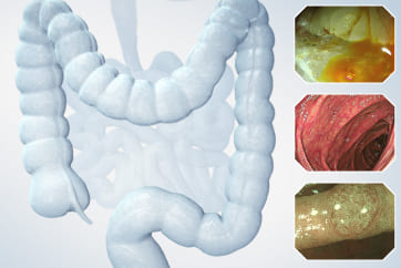
Case: A man in his 60s underwent a colonoscopy after a positive FIT test. He was found to have a cecal polyp and referred for endoscopic resection
Site of lesion: Colon/Appendiceal orifice
Patient information: M, 60s
Medical history: Melena
Other remarks: Family history of colon cancer in his father.
Past medical history: dyslipidemia, renal stones, osteoarthritis, L4-L5 decompression/fusion
1. 0-Is + IIa lesion - cecum
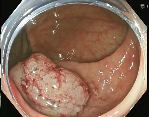
TXI 1
2. 0-Is + IIa - cecum
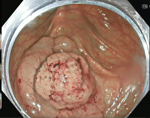
TXI 1
3. 0-Is + IIa - cecum
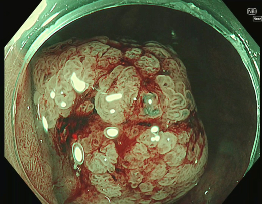
NBI + near focus
4. 0-Is + IIa - cecum
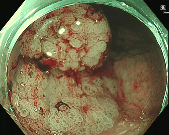
NBI + near focus
5. pEMR of IIa component
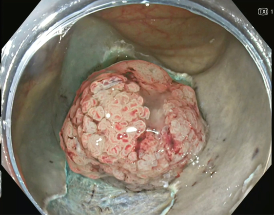
TXI 1
6. uEMR of IIa component
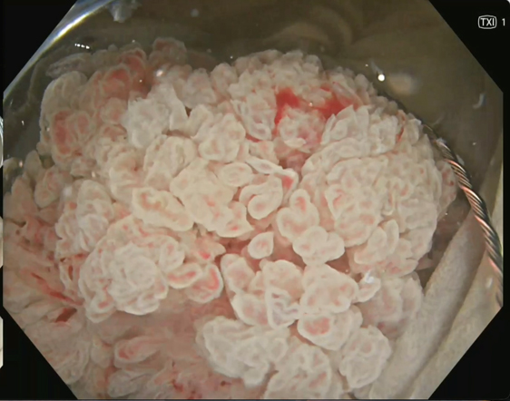
TXI 1
7. Post-EMR Defect
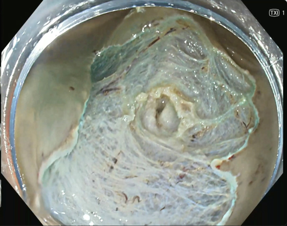
TXI 1
8. Appendiceal Orifice Post-EMR
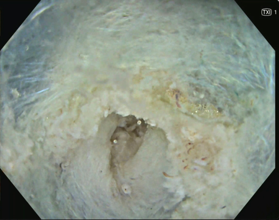
TXI 1
9. Appendiceal Orifice Pre-FTRD
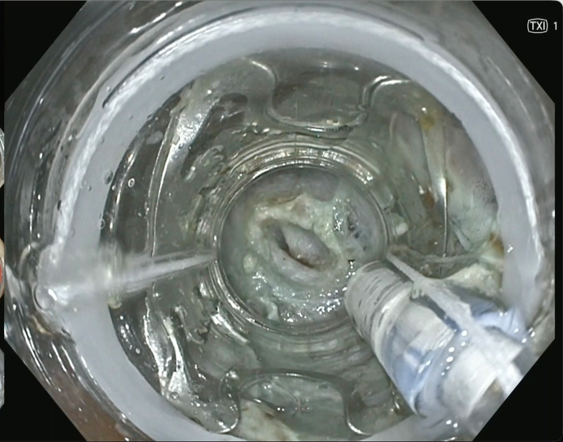
TXI 1
10. FTRD Clip Deployment
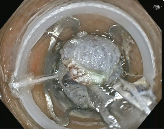
TXI 1
11. Post-FTRD Defect
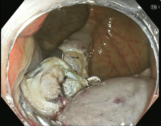
TXI 1
12. Specimen
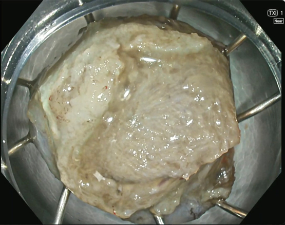
TXI 1 + Near Focus
Case Video
Overall Comment
Hybrid hot snare EMR and full-thickness resection (FTRD) of an appendiceal orifice lesion
A 30mm LST-G-M lesion in the cecum, involving the appendiceal orifice, was resected with a hybrid approach. The lesion was too large for en-bloc FTRD and involved the appendiceal orifice, which is unsafe for conventional EMR. Piecemeal hot snare EMR removed the cecal and peri-appendiceal component portions after submucosal lifting, leaving the residual appendiceal orifice component sized for FTRD. The FTRD was advanced to the residual lesion. The tissue was retracted into the cap with the grasper, the over-the-scope clip was deployed, and the tissue was snare-resected above the clip. The full-thickness specimen, including the proximal appendiceal stump, was retrieved and submitted separately from the EMR specimen so the margins could be assessed. The clip site showed a secure closure without visible perforation.
* Specifications, design and accessories are subject to change without any notice or obligation on the part of the manufacturer.
- Keyword
- Content Type