Disclaimer
The techniques and clinical opinions presented in this material reflect the personal experience and professional judgment of the healthcare professional and do not necessarily represent the views of Olympus. This material is intended for healthcare professionals only. Users should always refer to the applicable Instructions for Use (IFU) and use Olympus products in accordance with the approved indications and local regulatory requirements. The healthcare professional presenting this material has been engaged by Olympus and compensated at fair market value for their services.
Colorectal Case 35

Yasushi Sano 1.2
MD, PhD, FJGES, and ANBIIG 3
- Clinical Professor, Kansai Medical University Osaka, Japan
- Director & Chief of Gastrointestinal Center Sano Hospital, Kobe, Japan
- Secretary, Asian Novel Bio-Imaging and Intervention Group
Scope: CF-XZ1200I
Case: Detection
Site of lesion: Sigmoid colon
Patient information: M, 80s
Medical history: Screening
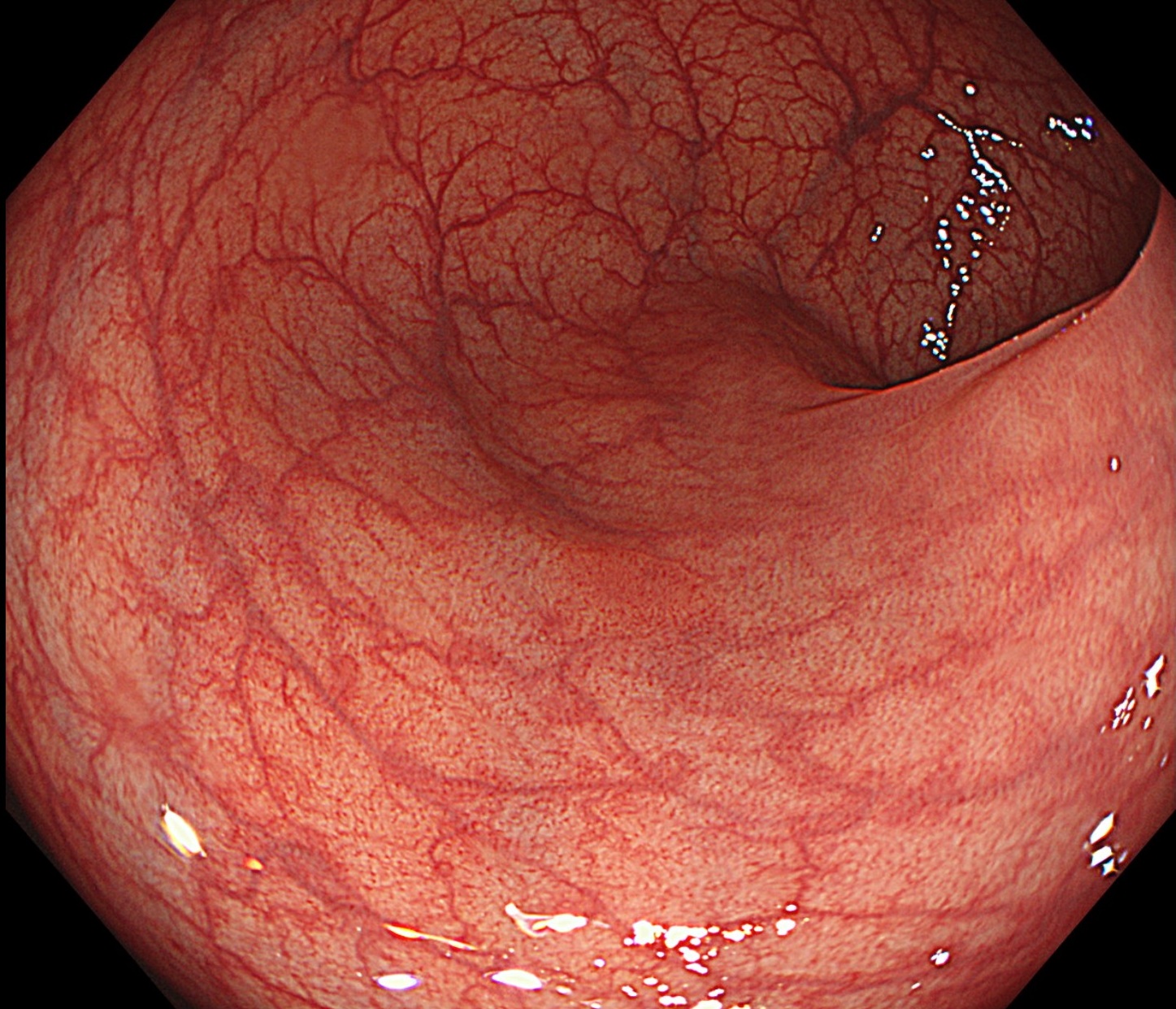
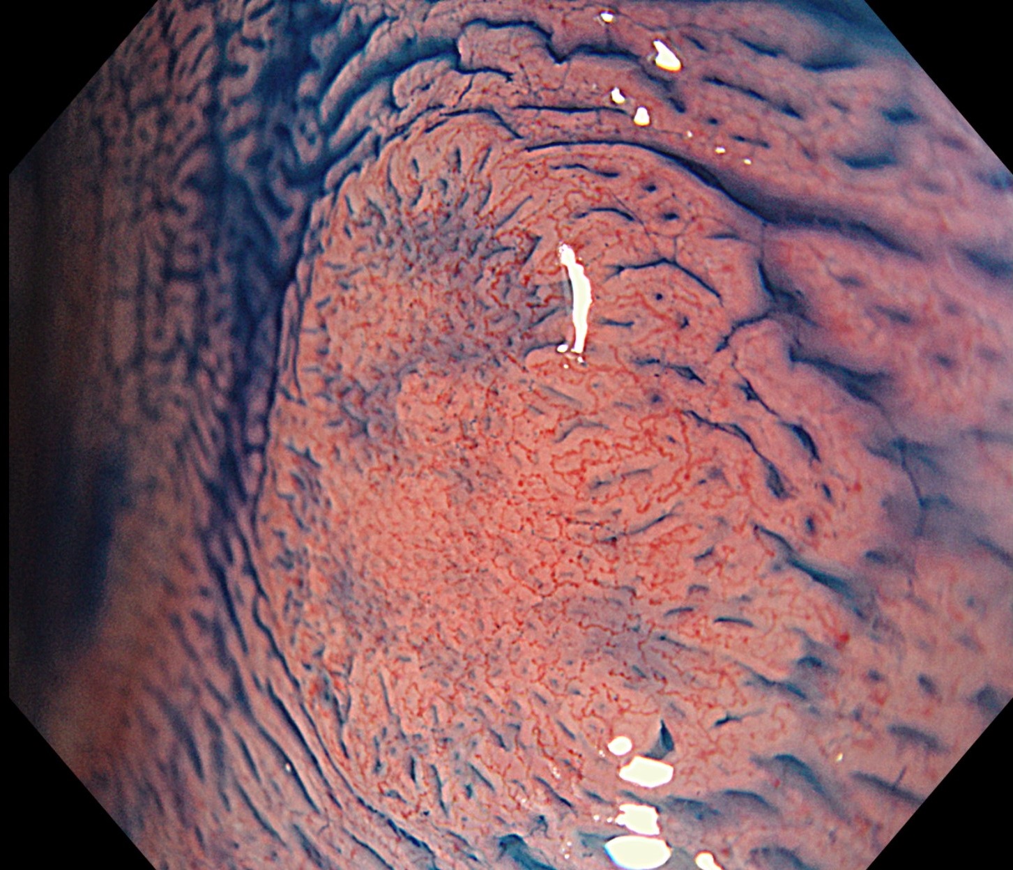
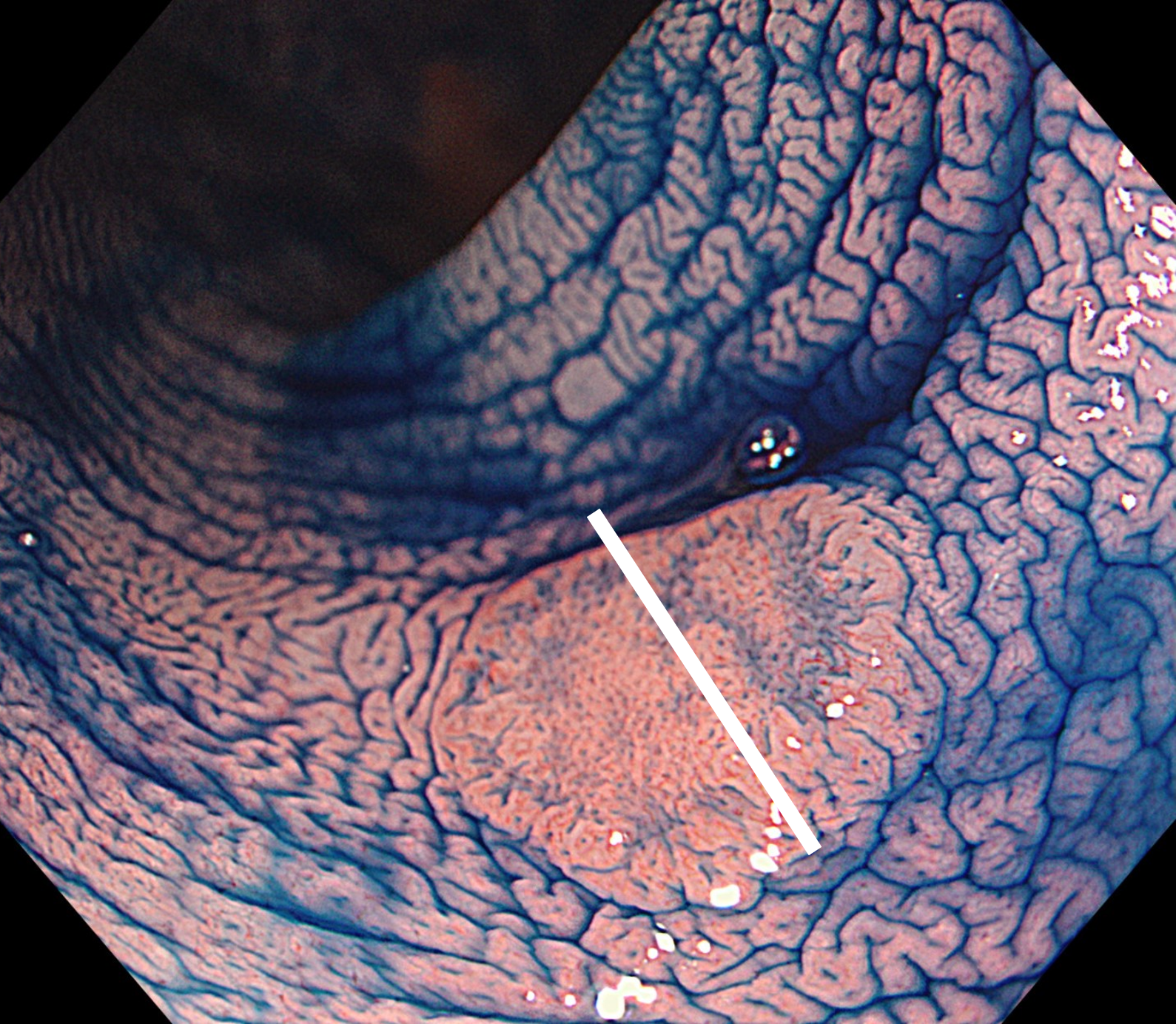
1. WLI
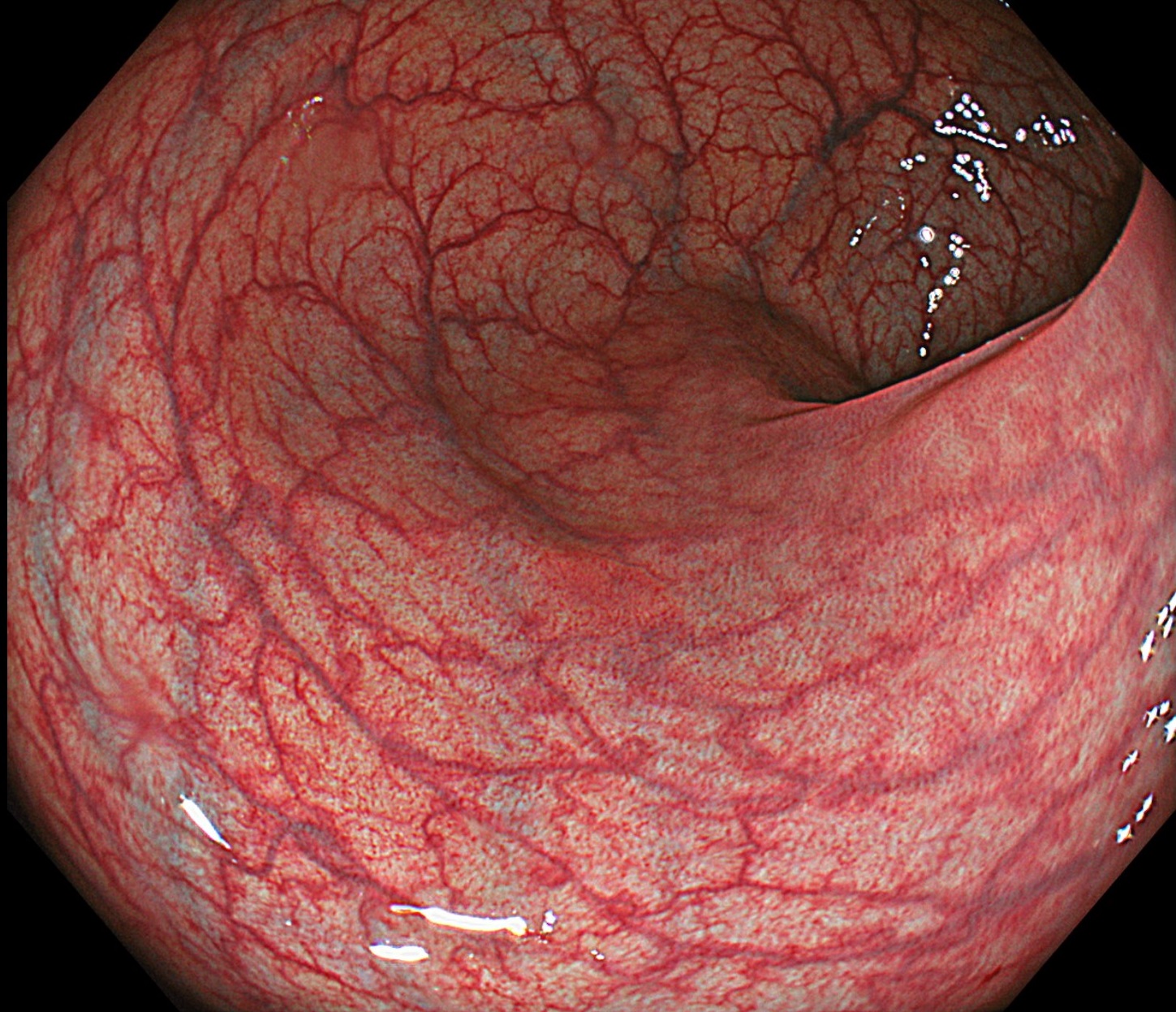
2. TXI
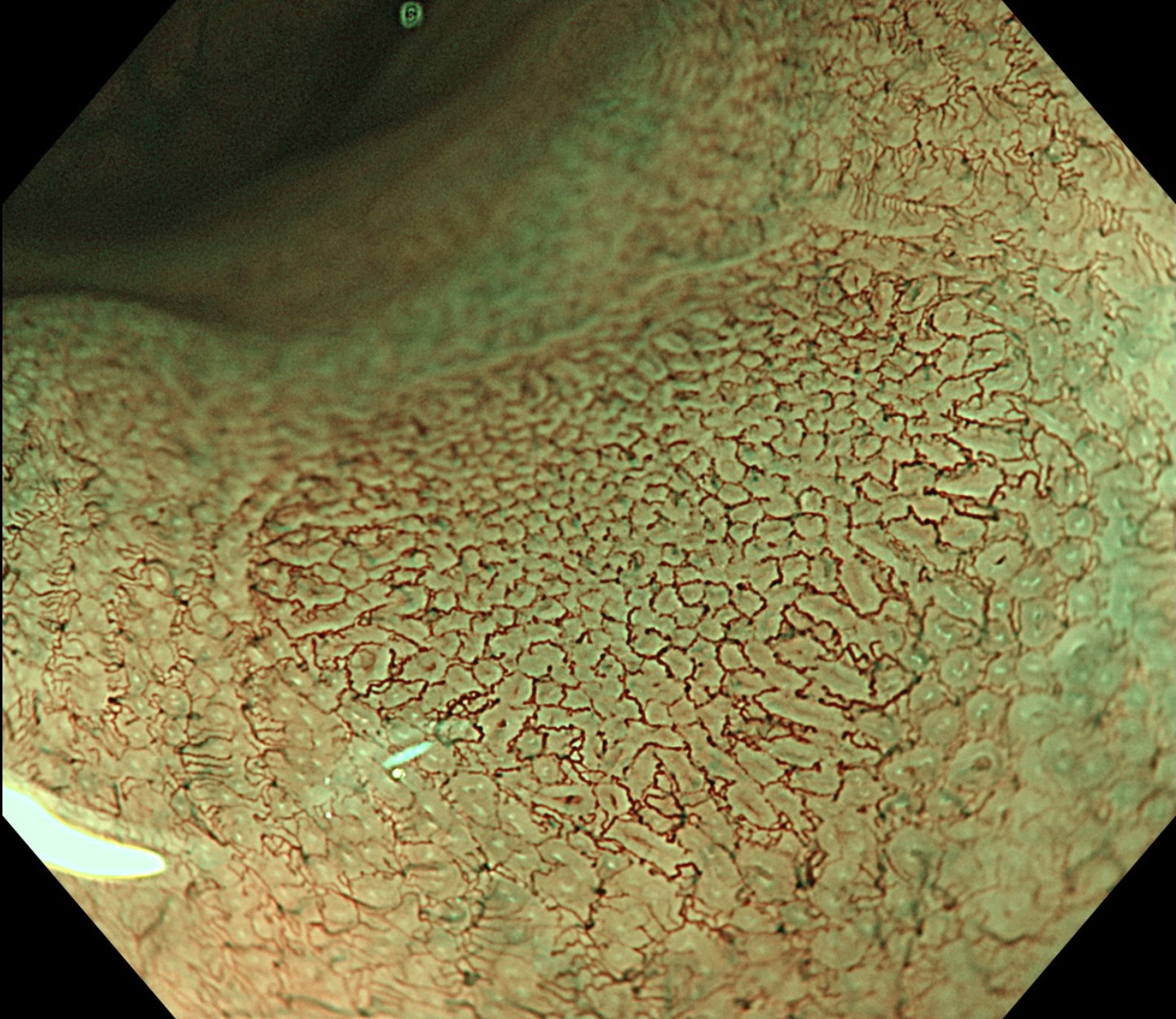
3. NBI
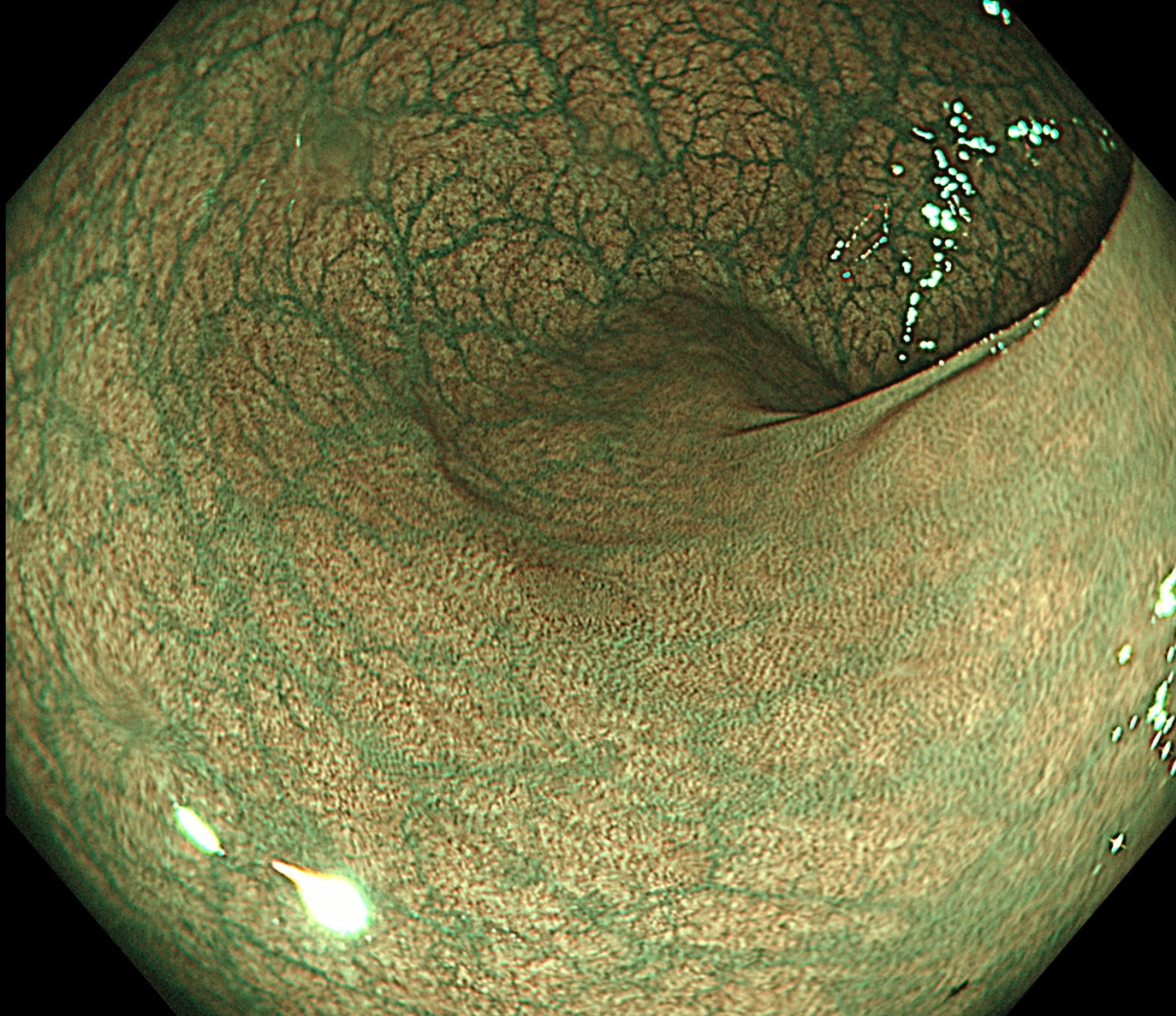
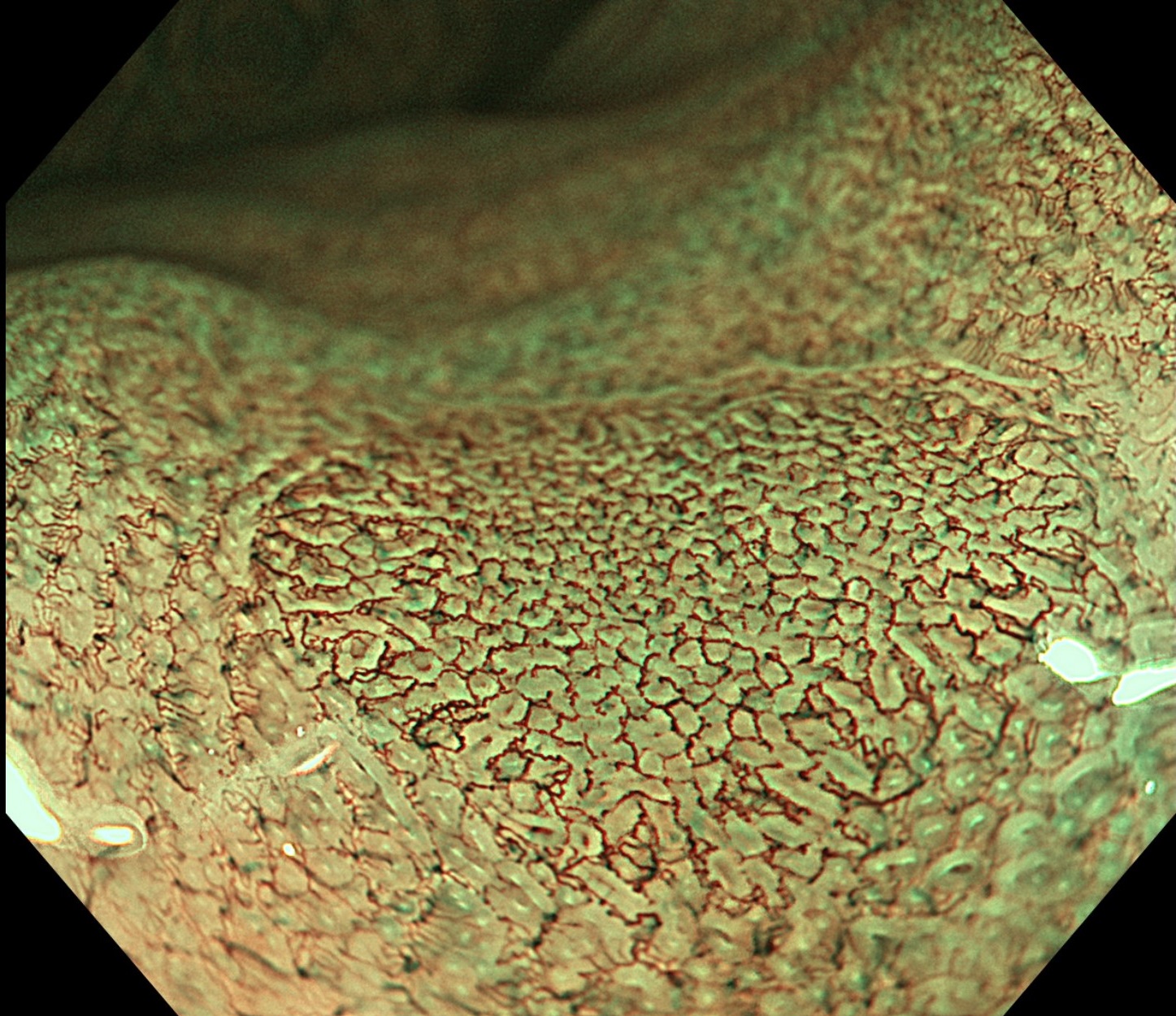
4. NBI
5. NBI
6. NBI+TXI
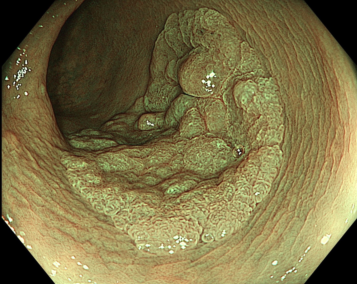
7. Chromoendoscopy
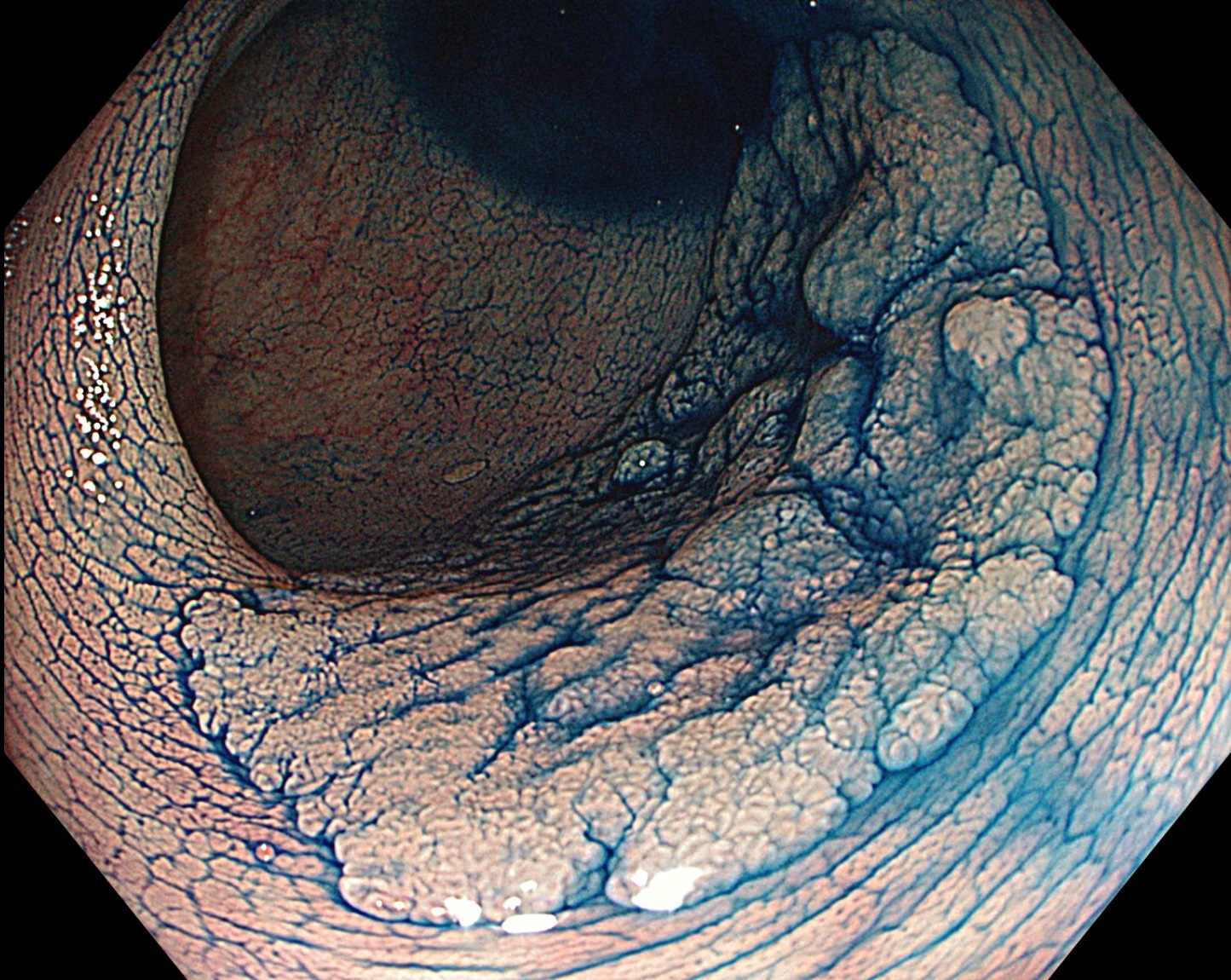
8. Chromoendoscopy
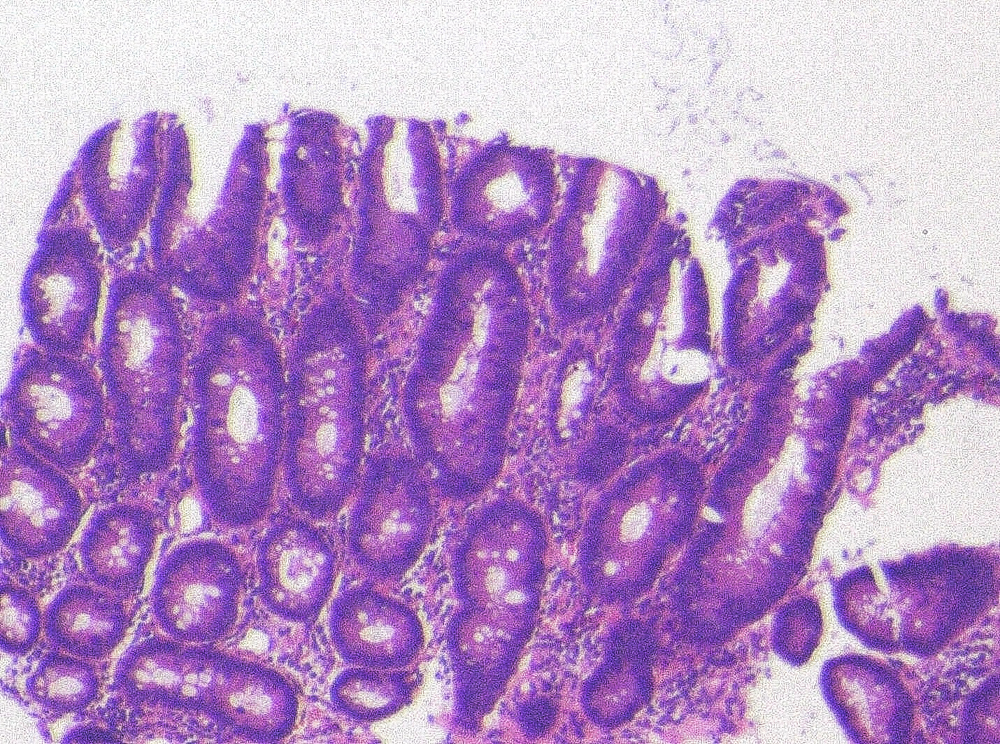
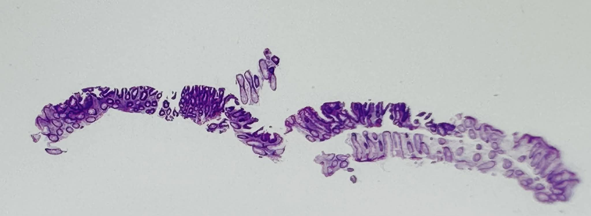
9. Pathological image
10. Pathological diagnosis
11. Pathological image
Case Video
Overall Comment
This case highlights the value of TXI and NBI-TXI for the detection and characterization of a diminutive colorectal adenoma. On white-light imaging, the lesion showed only subtle loss of vascular transparency and faint erythema. TXI enhanced these changes and improved lesion recognition. NBI identified the lesion as a brownish area, and NBI-TXI further increased its visibility. Magnifying observation demonstrated a regular vascular pattern and a Kudo type IIIL pit pattern, findings consistent with JNET Type 2A adenoma. Histopathology confirmed a tubular adenoma with moderate atypia.
* Specifications, design and accessories are subject to change without any notice or obligation on the part of the manufacturer.


